Explore
 Validate
 Learn
Learn Western blot
Antibody data
- Antibody Data
- Antigen structure
- References [0]
- Comments [0]
- Validations
- Western blot [2]
- Immunocytochemistry [1]
- Immunohistochemistry [1]
- Flow cytometry [1]
Submit
Validation data
Reference
Comment
Report error
- Product number
- TA325085 - Provider product page

- Provider
- OriGene
- Product name
- Rabbit polyclonal PIN1 Antibody (Center)
- Antibody type
- Polyclonal
- Description
- Rabbit polyclonal PIN1 Antibody (Center)
- Host
- Rabbit
- Conjugate
- Unconjugated
- Epitope
- PIN1
- Isotype
- IgG
- Antibody clone number
- NULL
- Vial size
- 400 µl
- Concentration
- 0.423 mg/ml
No comments: Submit comment
Supportive validation
- Submitted by
- OriGene (provider)
- Main image
- Experimental details
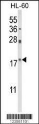
- Western blot analysis of PIN1 Antibody (Center) (Cat. #TA325085) in HL-60 cell line lysates (35ug/lane). PIN1 (arrow) was detected using the purified Pab.
- Validation comment
- WB

- Submitted by
- OriGene (provider)
- Main image

- Experimental details
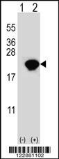
- Western blot analysis of PIN1 (arrow) using rabbit polyclonal PIN1 Antibody (Center) (Cat. #TA325085). 293 cell lysates (2 ug/lane) either nontransfected (Lane 1) or transiently transfected (Lane 2) with the PIN1 gene.
- Validation comment
- WB
Supportive validation
- Submitted by
- OriGene (provider)
- Main image
- Experimental details
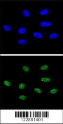
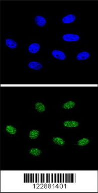
- Confocal immunofluorescent analysis of PIN1 Antibody (Center) (Cat. #TA325085) with 293 cell followed by Alexa Fluor?? 488-conjugated goat anti-rabbit lgG (green).DAPI was used to stain the cell nuclear (blue).
- Validation comment
- IF
Supportive validation
- Submitted by
- OriGene (provider)
- Main image
- Experimental details
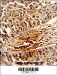
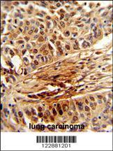
- Formalin-fixed and paraffin-embedded human lung carcinoma reacted with PIN1 Antibody (Center), which was peroxidase-conjugated to the secondary antibody, followed by DAB staining. This data demonstrates the use of this antibody for immunohistochemistry; clinical relevance has not been evaluated.
- Validation comment
- IHC
Supportive validation
- Submitted by
- OriGene (provider)
- Main image
- Experimental details
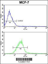
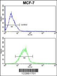
- PIN1 Antibody (Center) (Cat.#TA325085) FC analysis of MCF-7 cells (bottom histogram) compared to a negative control cell (top histogram). FITC-conjugated goat-anti-rabbit secondary antibodies were used for the analysis.
- Validation comment
- FC