Explore
 Validate
 Learn
Learn Western blot
 Immunoprecipitation
Antibody data
- Antibody Data
- Antigen structure
- References [6]
- Comments [0]
- Validations
- Western blot [1]
- Immunocytochemistry [1]
- Immunohistochemistry [1]
Submit
Validation data
Reference
Comment
Report error
- Product number
- MAB2294 - Provider product page

- Provider
- R&D Systems
- Product name
- Human/Mouse Pin1 Antibody
- Antibody type
- Monoclonal
- Description
- Protein A or G purified from hybridoma culture supernatant. Detects human and mouse Pin1 in Western blots.
- Reactivity
- Human, Mouse
- Host
- Mouse
- Conjugate
- Unconjugated
- Antigen sequence
Q13526- Isotype
- IgG
- Antibody clone number
- 257417
- Vial size
- 100 ug
- Concentration
- LYOPH
- Storage
- Use a manual defrost freezer and avoid repeated freeze-thaw cycles. 12 months from date of receipt, -20 to -70 °C as supplied. 1 month, 2 to 8 °C under sterile conditions after reconstitution. 6 months, -20 to -70 °C under sterile conditions after reconstitution.
Submitted references TLR-7 Stress Signaling in Differentiating and Mature Eosinophils Is Mediated by the Prolyl Isomerase Pin1.
The peptidyl-prolyl isomerase Pin1 up-regulation and proapoptotic function in dopaminergic neurons: relevance to the pathogenesis of Parkinson disease.
A distinct role for Pin1 in the induction and maintenance of pluripotency.
Protein Never in Mitosis Gene A Interacting-1 (PIN1) regulates degradation of inducible nitric oxide synthase in endothelial cells.
The peptidyl-isomerase Pin1 regulates p27kip1 expression through inhibition of Forkhead box O tumor suppressors.
Pin1 regulates TGF-beta1 production by activated human and murine eosinophils and contributes to allergic lung fibrosis.
Shen ZJ, Hu J, Kashi V, Bochkov YA, Gern JE, Malter JS
Journal of immunology (Baltimore, Md. : 1950) 2018 Dec 15;201(12):3503-3513
Journal of immunology (Baltimore, Md. : 1950) 2018 Dec 15;201(12):3503-3513
The peptidyl-prolyl isomerase Pin1 up-regulation and proapoptotic function in dopaminergic neurons: relevance to the pathogenesis of Parkinson disease.
Ghosh A, Saminathan H, Kanthasamy A, Anantharam V, Jin H, Sondarva G, Harischandra DS, Qian Z, Rana A, Kanthasamy AG
The Journal of biological chemistry 2013 Jul 26;288(30):21955-71
The Journal of biological chemistry 2013 Jul 26;288(30):21955-71
A distinct role for Pin1 in the induction and maintenance of pluripotency.
Nishi M, Akutsu H, Masui S, Kondo A, Nagashima Y, Kimura H, Perrem K, Shigeri Y, Toyoda M, Okayama A, Hirano H, Umezawa A, Yamamoto N, Lee SW, Ryo A
The Journal of biological chemistry 2011 Apr 1;286(13):11593-603
The Journal of biological chemistry 2011 Apr 1;286(13):11593-603
Protein Never in Mitosis Gene A Interacting-1 (PIN1) regulates degradation of inducible nitric oxide synthase in endothelial cells.
Liu T, Huang Y, Likhotvorik RI, Keshvara L, Hoyt DG
American journal of physiology. Cell physiology 2008 Sep;295(3):C819-27
American journal of physiology. Cell physiology 2008 Sep;295(3):C819-27
The peptidyl-isomerase Pin1 regulates p27kip1 expression through inhibition of Forkhead box O tumor suppressors.
Brenkman AB, de Keizer PL, van den Broek NJ, van der Groep P, van Diest PJ, van der Horst A, Smits AM, Burgering BM
Cancer research 2008 Sep 15;68(18):7597-605
Cancer research 2008 Sep 15;68(18):7597-605
Pin1 regulates TGF-beta1 production by activated human and murine eosinophils and contributes to allergic lung fibrosis.
Shen ZJ, Esnault S, Rosenthal LA, Szakaly RJ, Sorkness RL, Westmark PR, Sandor M, Malter JS
The Journal of clinical investigation 2008 Feb;118(2):479-90
The Journal of clinical investigation 2008 Feb;118(2):479-90
No comments: Submit comment
Supportive validation
- Submitted by
- R&D Systems (provider)
- Main image
- Experimental details
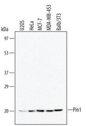
- Detection of Human/Mouse Pin1 by Western Blot. Western blot shows lysates of Balb/3T3 mouse embryonic fibroblast cell line, U2OS human osteosarcoma cell line, HeLa human cervical epithelial carcinoma cell line, MCF-7 human breast cancer cell line, and MDA-MB-453 human breast cancer cell line. PVDF membrane was probed with 0.5 µg/mL of Mouse Anti-Human/Mouse Pin1 Monoclonal Antibody (Catalog # MAB2294) followed by HRP-conjugated Anti-Mouse IgG Secondary Antibody (Catalog # HAF007). A specific band was detected for Pin1 at approximately 20 kDa (as indicated). This experiment was conducted under reducing conditions and using Immunoblot Buffer Group 1.
Supportive validation
- Submitted by
- R&D Systems (provider)
- Main image

- Experimental details
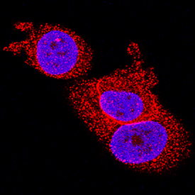
- Pin1 in MCF-7 Human Cell Line. Pin1 was detected in immersion fixed MCF-7 human breast cancer cell line using Mouse Anti-Human/Mouse Pin1 Monoclonal Antibody (Catalog # MAB2294) at 8 µg/mL for 3 hours at room temperature. Cells were stained using the NorthernLights™ 557-conjugated Anti-Mouse IgG Secondary Antibody (red; Catalog # NL007) and counterstained with DAPI (blue). Specific staining was localized to nuclei and cytoplasm. View our protocol for Fluorescent ICC Staining of Cells on Coverslips.
Supportive validation
- Submitted by
- R&D Systems (provider)
- Main image
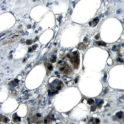
- Experimental details
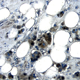
- Pin1 in Human Breast Cancer Tissue. Pin1 was detected in immersion fixed paraffin-embedded sections of human breast cancer tissue using Mouse Anti-Human/Mouse Pin1 Monoclonal Antibody (Catalog # MAB2294) at 25 µg/mL overnight at 4 °C. Tissue was stained using the Anti-Mouse HRP-DAB Cell & Tissue Staining Kit (brown; Catalog # CTS002) and counterstained with hematoxylin (blue). Specific labeling was localized to the cytoplasm of epithelial cells. View our protocol for Chromogenic IHC Staining of Paraffin-embedded Tissue Sections.