Explore
 Validate
 Learn
Learn Western blot
Antibody data
- Antibody Data
- Antigen structure
- References [0]
- Comments [0]
- Validations
- Western blot [1]
- Immunocytochemistry [1]
Submit
Validation data
Reference
Comment
Report error
- Product number
- PA5-65442 - Provider product page

- Provider
- Invitrogen Antibodies
- Product name
- Reelin Polyclonal Antibody
- Antibody type
- Polyclonal
- Antigen
- Recombinant full-length protein
- Description
- Immunogen sequence: TFCEPYGPREL ITTGLNTTTA SVLQFSIGSG SCRFSYSDPS IIVLYAKNNS ADWIQLEKIR APSNVSTIIH ILYLPEDAKG ENVQFQWKQE NLRVGEVYE
- Concentration
- 0.2 mg/mL
No comments: Submit comment
Supportive validation
- Submitted by
- Invitrogen Antibodies (provider)
- Main image
- Experimental details
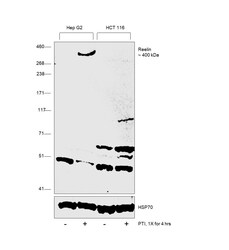
- Western Blot was performed using Anti-Reelin Polyclonal Antibody (Product # PA5-65442) and a 400 kDa band corresponding to Reelin was observed across Hep G2 treated with PTI when compared to HCT 116. Whole cell extracts (30 µg lysate) of Hep G2 (Lane 1), Hep G2 treated with PTI (1X, for 4 hrs) (Lane 2), HCT 116 (Lane 3), HCT 116 treated with PTI (1X, for 4 hrs) (Lane 4) were electrophoresed using NuPAGE™ 3-8% Tris-Acetate Protein Gel (Product # EA0378BOX). Resolved proteins were then transferred onto a Nitrocellulose membrane (Product # IB23001) by iBlot® 2 Dry Blotting System (Product # IB21001). The Blot was probed with the primary antibody (1:1000 dilution) and detected by chemiluminescence with Goat anti-Rabbit IgG (H+L) Superclonal™ Recombinant Secondary Antibody, HRP (Product # A27036, 1:6000 dilution) using the iBright FL 1000 (Product # A32752). Chemiluminescent detection was performed using SuperSignal™ West Dura Extended Duration Substrate (Product # 34076). Few uncharacterized bands were observed between 50-70 kDa.

Supportive validation
- Submitted by
- Invitrogen Antibodies (provider)
- Main image
- Experimental details
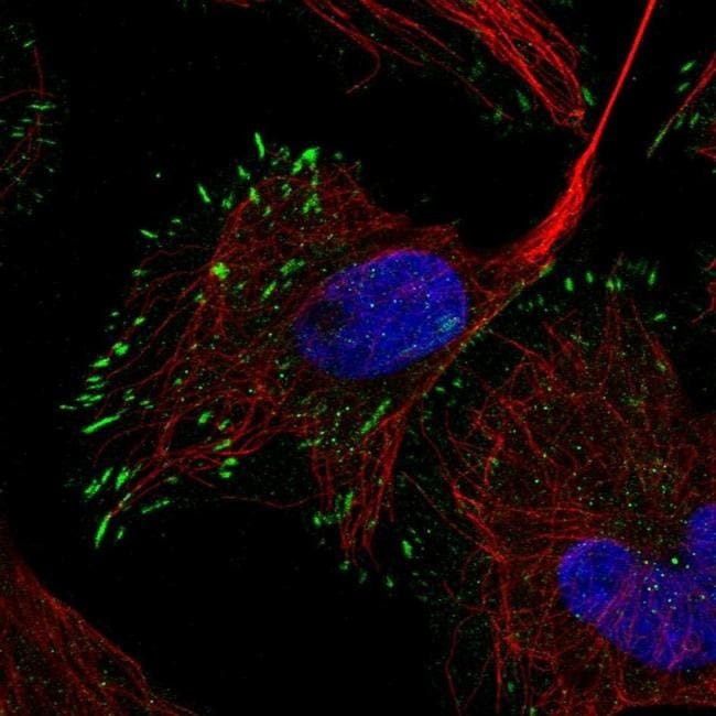
- Immunofluorescent staining of Reelin in human cell line U-2 OS shows localization to focal adhesion sites. Samples were probed using a Reelin Polyclonal Antibody (Product # PA5-65442).