Explore
 Validate
 Learn
Learn433900
antibody from Invitrogen Antibodies
Targeting: RXRA
NR2B1
 Western blot
 ELISA
ELISA Immunocytochemistry Immunoprecipitation Immunohistochemistry Gel shift Chromatin Immunoprecipitation Other assay
Antibody data
- Antibody Data
- Antigen structure
- References [1]
- Comments [0]
- Validations
- Immunocytochemistry [3]
- Immunoprecipitation [1]
- Immunohistochemistry [2]
- Other assay [1]
Submit
Validation data
Reference
Comment
Report error
- Product number
- 433900 - Provider product page

- Provider
- Invitrogen Antibodies
- Product name
- RXRA Monoclonal Antibody (K8508)
- Antibody type
- Monoclonal
- Antigen
- Recombinant full-length protein
- Description
- This antibody specifically recognizes human RXR alpha and cross reacts with mouse and rat RXR alpha. This antibody does not recognize human RXR beta and gamma.
- Reactivity
- Human, Mouse, Rat
- Host
- Mouse
- Isotype
- IgG
- Antibody clone number
- K8508
- Vial size
- 100 μL
- Concentration
- 1 mg/mL
- Storage
- Maintain refrigerated at 2-8°C for up to 1 month. For long term storage store at -20°C
Submitted references Macrophages activated by hepatitis B virus have distinct metabolic profiles and suppress the virus via IL-1β to downregulate PPARα and FOXO3.
Li Y, Zhu Y, Feng S, Ishida Y, Chiu TP, Saito T, Wang S, Ann DK, Ou JJ
Cell reports 2022 Jan 25;38(4):110284
Cell reports 2022 Jan 25;38(4):110284
No comments: Submit comment
Supportive validation
- Submitted by
- Invitrogen Antibodies (provider)
- Main image
- Experimental details
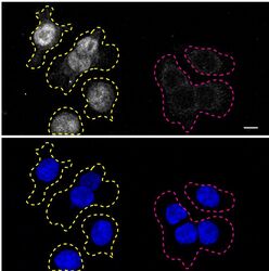
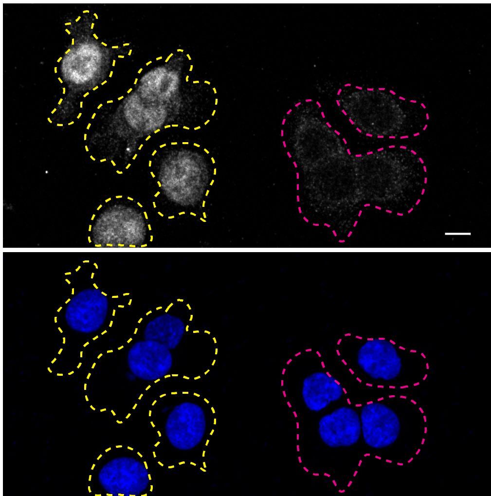
- Immunofluorescence of RXR-alpha was performed using HCT116 wild-type and RXR-alpha KO cells that were transfected with a green or a far-red fluorescent dye, respectively. Post-transfection, WT and KO cells were mixed and plated to a 1:1 ratio on coverslips as a mosaic and incubated for 24 hrs. Cells were fixed in 4% PFA (in PBS) or methanol for 15 min; cells were permeabilized with 0.1% Triton X-100 for 10 min at RT and blocked with PBS with 5% BSA, 5% goat serum, and 0.01% Triton X-100 for 30 min. Cells were stained with the RXR-alpha monoclonal antibody (Product # 433900) at a 1:1,000 dilution overnight at 4°C. Secondary antibody incubation was performed using 1 µg/mL of Goat anti-Mouse IgG (H+L) Highly Cross-Adsorbed Secondary Antibody, Alexa Fluor 555 antibody (Product # A21424) together with DAPI for 1 hr. Imaging was performed with a 40X oil objective and analysis was performed using Image J. Cell image represents a single focal plane; WT and KO cells are outlined with a yellow (WT) or magenta (KO) dashed line. Data courtesy of YCharOS Inc., an open science company with the mission of characterizing commercially available antibodies using knockout validation.
- Submitted by
- Invitrogen Antibodies (provider)
- Main image

- Experimental details
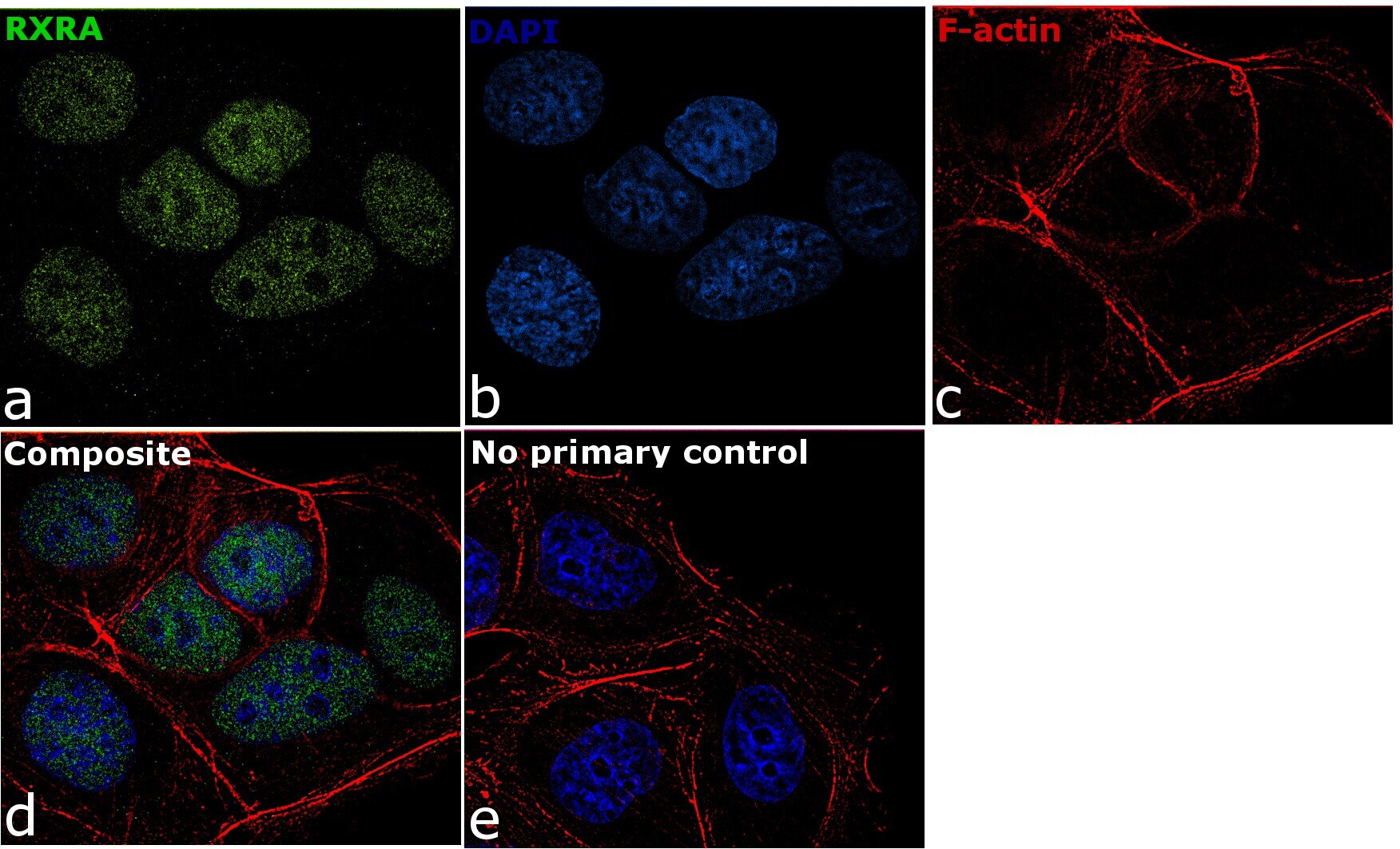
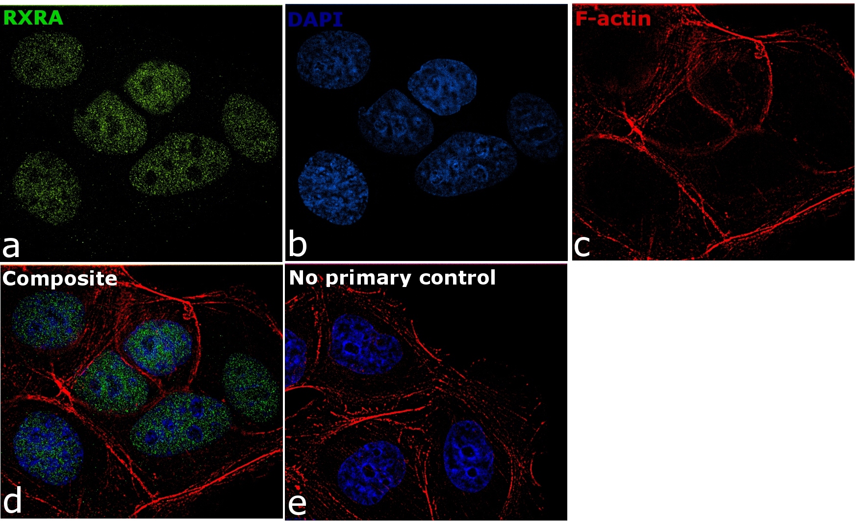
- Immunofluorescence analysis of RXRA was performed using 70% confluent log phase MCF7 cells. The cells were fixed with 4% paraformaldehyde for 10 minutes, permeabilized with 0.1% Triton™ X-100 for 10 minutes, and blocked with 1% BSA for 1 hour at room temperature. The cells were labeled with RXRA Monoclonal Antibody (Product # 433900) at 5 µg/mL in 0.1% BSA and incubated overnight at 4 degree and then labeled with Goat anti-Mouse IgG (H+L) Superclonal™ Secondary Antibody, Alexa Fluor® 488 conjugate (Product # A28175) at a dilution of 1:2000 for 45 minutes at room temperature (Panel a: green). Nuclei (Panel b: blue) were stained with SlowFade® Gold Antifade Mountant with DAPI (Product # S36938). F-actin (Panel c: red) was stained with Rhodamine Phalloidin (Product # R415, 1:300). Panel d represents the merged image showing nuclear localization. Panel e represents control cells with no primary antibody to assess background. The images were captured at 60X magnification.
- Submitted by
- Invitrogen Antibodies (provider)
- Main image

- Experimental details
- Immunofluorescence analysis of RXRA was performed using 70% confluent log phase MCF7 cells. The cells were fixed with 4% paraformaldehyde for 10 minutes, permeabilized with 0.1% Triton™ X-100 for 10 minutes, and blocked with 1% BSA for 1 hour at room temperature. The cells were labeled with RXRA Monoclonal Antibody (Product # 433900) at 5 µg/mL in 0.1% BSA and incubated overnight at 4 degree and then labeled with Goat anti-Mouse IgG (H+L) Superclonal™ Secondary Antibody, Alexa Fluor® 488 conjugate (Product # A28175) at a dilution of 1:2000 for 45 minutes at room temperature (Panel a: green). Nuclei (Panel b: blue) were stained with SlowFade® Gold Antifade Mountant with DAPI (Product # S36938). F-actin (Panel c: red) was stained with Rhodamine Phalloidin (Product # R415, 1:300). Panel d represents the merged image showing nuclear localization. Panel e represents control cells with no primary antibody to assess background. The images were captured at 60X magnification.
Supportive validation
- Submitted by
- Invitrogen Antibodies (provider)
- Main image
- Experimental details
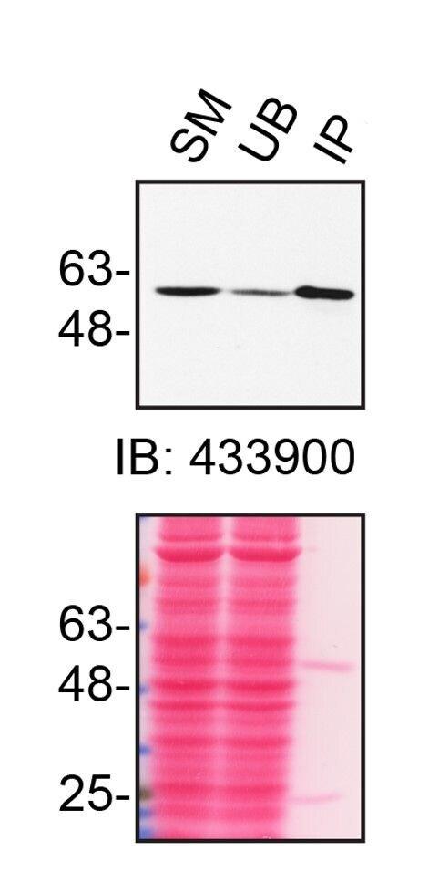
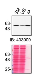
- Immunoprecipitation of RXR-alpha was performed on HCT116 cell lysates. Antibody-bead conjugates were prepared by adding 1 µg of RXR-alpha monoclonal antibody (Product # 433900) with 30 µL of protein G-Sepharose beads and rocked overnight at 4°C. 1 mg of lysate was incubated with an antibody-bead conjugate for 2 hours at 4°C. Following centrifugation and multiple washes, 10% starting material (SM), 10% unbound fraction (UB) and immunoprecipitated fraction (IP) were processed for immunoblot using the same antibody. Ponceau stained transfer of blot is shown. Data courtesy of YCharOS Inc., an open science company with the mission of characterizing commercially available antibodies using knockout validation.

Supportive validation
- Submitted by
- Invitrogen Antibodies (provider)
- Main image
- Experimental details
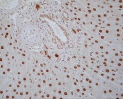
- Immunohistochemistry analysis of RXR alpha in paraffin-embedded rat liver tissue. The sample was incubated in RXR alpha monoclonal antibody (Product # 433900) at a dilution of 3~20 µg/mL. Section was fixed with 10% formalin-fixed in immersion, activated by autoclave at 121°C for 15 minutes, and blocked with 3% H2O2 in methanol.

- Submitted by
- Invitrogen Antibodies (provider)
- Main image
- Experimental details
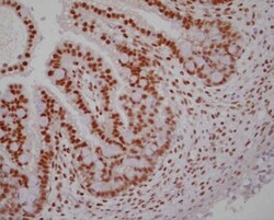
- Immunohistochemistry analysis of RXR alpha in paraffin-embedded rat embryonic intestine. The sample was incubated in RXR alpha monoclonal antibody (Product # 433900) at a dilution of 3~20 μg/mL. Section was fixed with 10% formalin-fixed in immersion, activated by autoclave at 121°C for 15 minutes, and blocked with 3% H2O2 in methanol.

Supportive validation
- Submitted by
- Invitrogen Antibodies (provider)
- Main image

- Experimental details
- Immunoprecipitation of RXR-alpha was performed on HCT116 cell lysates. Antibody-bead conjugates were prepared by adding 1 µg of RXR-alpha monoclonal antibody (Product # 433900) with 30 µL of protein G-Sepharose beads and rocked overnight at 4°C. 1 mg of lysate was incubated with an antibody-bead conjugate for 2 hours at 4°C. Following centrifugation and multiple washes, 10% starting material (SM), 10% unbound fraction (UB) and immunoprecipitated fraction (IP) were processed for immunoblot using the same antibody. Ponceau stained transfer of blot is shown. Data courtesy of YCharOS Inc., an open science company with the mission of characterizing commercially available antibodies using knockout validation.