Explore
 Validate
 Learn
Learn Western blot
Antibody data
- Antibody Data
- Antigen structure
- References [0]
- Comments [0]
- Validations
- Western blot [2]
- Immunocytochemistry [6]
- Immunoprecipitation [1]
- Immunohistochemistry [4]
- Other assay [1]
Submit
Validation data
Reference
Comment
Report error
- Product number
- MA5-34806 - Provider product page

- Provider
- Invitrogen Antibodies
- Product name
- RXRA Recombinant Rabbit Monoclonal Antibody (JG99-38)
- Antibody type
- Monoclonal
- Antigen
- Recombinant full-length protein
- Description
- Positive Control: MCF-7, rat liver tissue, human esophagus tissue.
- Reactivity
- Human
- Host
- Rabbit
- Isotype
- IgG
- Antibody clone number
- JG99-38
- Vial size
- 100 μL
- Concentration
- 1 mg/mL
- Storage
- -20°C, Avoid Freeze/Thaw Cycles, store in dark
No comments: Submit comment
Supportive validation
- Submitted by
- Invitrogen Antibodies (provider)
- Main image
- Experimental details
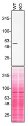
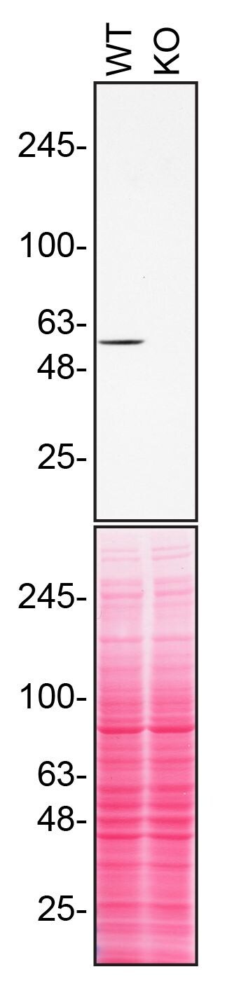
- Western blot of RXR-alpha was performed by loading 100 µg of WT (lane 1) and RXR-alpha CRISPR KO (lane 2) HCT116 cell lysates in RIPA buffer onto a 4-15% gradient polyacrylamide gel. Proteins on the blots were visualized with Ponceau staining (below immunoblot). Proteins were transferred to nitrocellulose membrane and blocked in 5% milk for 1 hr. RXR-alpha was detected at approximately 50 kDa using an RXR-alpha recombinant monoclonal antibody (Product # MA5-34806) at a dilution of 1:500 in 5% BSA in TBS with 0.1% Tween 20 (TBST) overnight at 4°C. The peroxidase-conjugated secondary antibody (Product # 65-6120) was diluted to 0.2 µg/mL in TBST with 5% milk for 1 hr. Chemiluminescent detection was performed using Pierce ECL Western Blotting Substrate (Product # 32106). Data courtesy of YCharOS Inc., an open science company with the mission of characterizing commercially available antibodies using knockout validation.
- Submitted by
- Invitrogen Antibodies (provider)
- Main image

- Experimental details
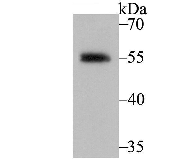
- Western blot analysis of Retinoid X Receptor alpha in MCF-7 cell. Samples were incubated with Retinoid X Receptor alpha monoclonal antibody (Product # MA5-34806), at a dilution of 1:5000.
Supportive validation
- Submitted by
- Invitrogen Antibodies (provider)
- Main image

- Experimental details
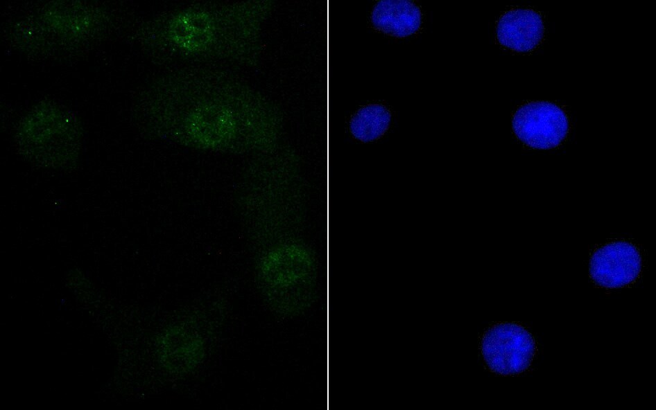
- Immunofluorescent analysis of Retinoid X Receptor alpha in HeLa cells (green). Samples were fixed in paraformaldehyde and permeabilised with 0.25% Triton X100/PBS, incubated with Retinoid X Receptor alpha monoclonal antibody (Product # MA5-34806), followed by DAPI (blue).

- Submitted by
- Invitrogen Antibodies (provider)
- Main image

- Experimental details
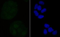
- Immunofluorescent analysis of Retinoid X Receptor alpha in A549 cells (green). Samples were fixed in paraformaldehyde and permeabilised with 0.25% Triton X100/PBS, incubated with Retinoid X Receptor alpha monoclonal antibody (Product # MA5-34806), followed by DAPI (blue).

- Submitted by
- Invitrogen Antibodies (provider)
- Main image
- Experimental details
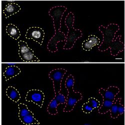
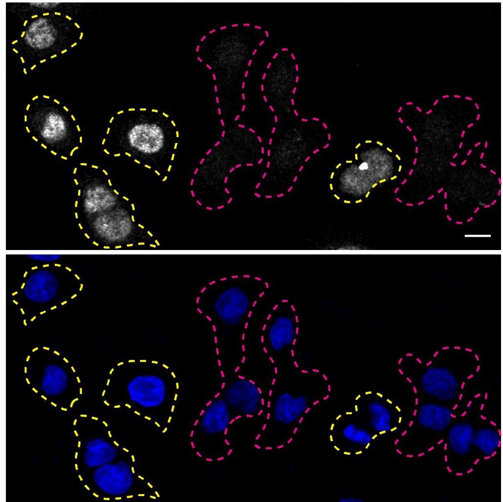
- Immunofluorescence of RXR-alpha was performed using HCT116 wild-type and RXR-alpha KO cells that were transfected with a green or a far-red fluorescent dye, respectively. Post-transfection, WT and KO cells were mixed and plated to a 1:1 ratio on coverslips as a mosaic and incubated for 24 hrs. Cells were fixed in 4% PFA (in PBS) or methanol for 15 min; cells were permeabilized with 0.1% Triton X-100 for 10 min at RT and blocked with PBS with 5% BSA, 5% goat serum, and 0.01% Triton X-100 for 30 min. Cells were stained with the RXR-alpha recombinant monoclonal antibody (Product # MA5-34806) at a 1:1,000 dilution overnight at 4°C. Secondary antibody incubation was performed using 1 µg/mL of Goat anti-Rabbit IgG (H+L) Highly Cross-Adsorbed Secondary Antibody, Alexa Fluor 555 antibody (Product # A21429) together with DAPI for 1 hr. Imaging was performed with a 40X oil objective and analysis was performed using Image J. Cell image represents a single focal plane; WT and KO cells are outlined with a yellow (WT) or magenta (KO) dashed line. Data courtesy of YCharOS Inc., an open science company with the mission of characterizing commercially available antibodies using knockout validation.

- Submitted by
- Invitrogen Antibodies (provider)
- Main image

- Experimental details
- Immunofluorescent analysis of Retinoid X Receptor alpha in A549 cells (green). Samples were fixed in paraformaldehyde and permeabilised with 0.25% Triton X100/PBS, incubated with Retinoid X Receptor alpha monoclonal antibody (Product # MA5-34806), followed by DAPI (blue).
- Submitted by
- Invitrogen Antibodies (provider)
- Main image
- Experimental details
- Immunofluorescent analysis of Retinoid X Receptor alpha in HeLa cells (green). Samples were fixed in paraformaldehyde and permeabilised with 0.25% Triton X100/PBS, incubated with Retinoid X Receptor alpha monoclonal antibody (Product # MA5-34806), followed by DAPI (blue).

- Submitted by
- Invitrogen Antibodies (provider)
- Main image

- Experimental details
- Immunofluorescence of RXR-alpha was performed using HCT116 wild-type and RXR-alpha KO cells that were transfected with a green or a far-red fluorescent dye, respectively. Post-transfection, WT and KO cells were mixed and plated to a 1:1 ratio on coverslips as a mosaic and incubated for 24 hrs. Cells were fixed in 4% PFA (in PBS) or methanol for 15 min; cells were permeabilized with 0.1% Triton X-100 for 10 min at RT and blocked with PBS with 5% BSA, 5% goat serum, and 0.01% Triton X-100 for 30 min. Cells were stained with the RXR-alpha recombinant monoclonal antibody (Product # MA5-34806) at a 1:1,000 dilution overnight at 4°C. Secondary antibody incubation was performed using 1 µg/mL of Goat anti-Rabbit IgG (H+L) Highly Cross-Adsorbed Secondary Antibody, Alexa Fluor 555 antibody (Product # A21429) together with DAPI for 1 hr. Imaging was performed with a 40X oil objective and analysis was performed using Image J. Cell image represents a single focal plane; WT and KO cells are outlined with a yellow (WT) or magenta (KO) dashed line. Data courtesy of YCharOS Inc., an open science company with the mission of characterizing commercially available antibodies using knockout validation.
Supportive validation
- Submitted by
- Invitrogen Antibodies (provider)
- Main image

- Experimental details
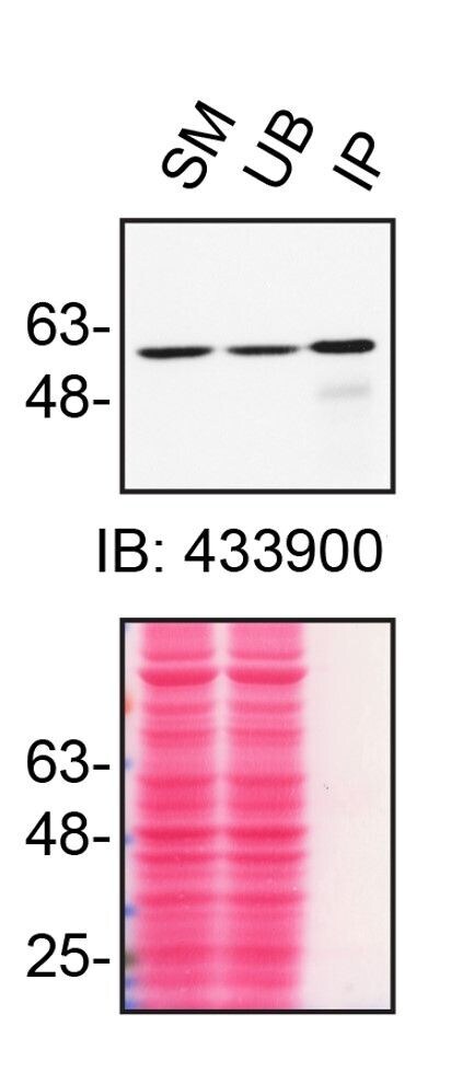
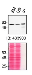
- Immunoprecipitation of RXR-alpha was performed on HCT116 cell lysates. Antibody-bead conjugates were prepared by adding 1 µg of RXR-alpha recombinant monoclonal antibody (Product # MA5-34806) with 30 µL of protein A-Sepharose beads and rocked overnight at 4°C. 1 mg of lysate was incubated with an antibody-bead conjugate for 2 hours at 4°C. Following centrifugation and multiple washes, 10% starting material (SM), 10% unbound fraction (UB) and immunoprecipitated fraction (IP) were processed for immunoblot using a different antibody (Product # 433900). Ponceau stained transfer of blot is shown. Data courtesy of YCharOS Inc., an open science company with the mission of characterizing commercially available antibodies using knockout validation.

Supportive validation
- Submitted by
- Invitrogen Antibodies (provider)
- Main image
- Experimental details
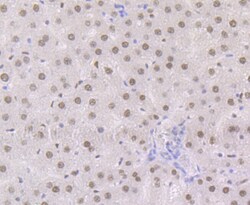
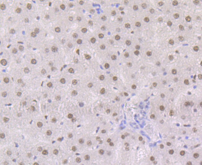
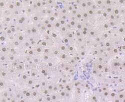
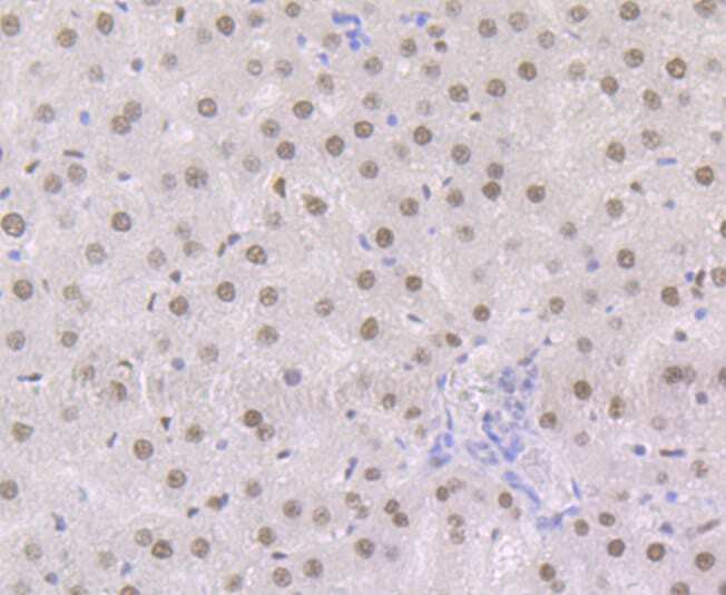
- Immunohistochemistry analysis of Retinoid X Receptor alpha in paraffin-embedded rat liver tissue. Samples were incubated with Retinoid X Receptor alpha monoclonal antibody (Product # MA5-34806), and followed by hematoxylin.
- Submitted by
- Invitrogen Antibodies (provider)
- Main image
- Experimental details
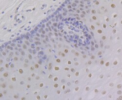
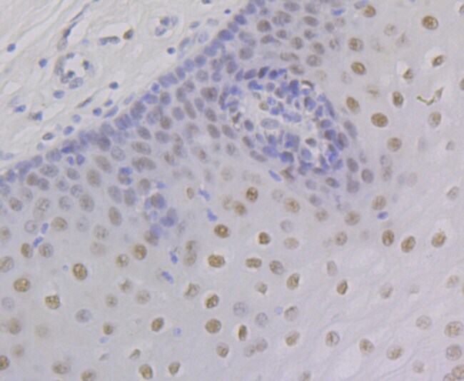
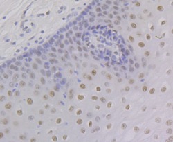
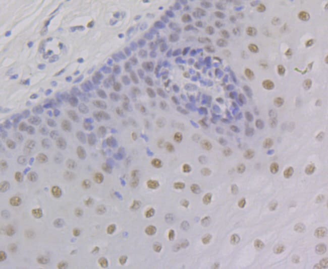
- Immunohistochemistry analysis of Retinoid X Receptor alpha in paraffin-embedded human esophagus tissue. Samples were incubated with Retinoid X Receptor alpha monoclonal antibody (Product # MA5-34806), and followed by hematoxylin.
- Submitted by
- Invitrogen Antibodies (provider)
- Main image
- Experimental details
- Immunohistochemistry analysis of Retinoid X Receptor alpha in paraffin-embedded rat liver tissue. Samples were incubated with Retinoid X Receptor alpha monoclonal antibody (Product # MA5-34806), and followed by hematoxylin.
- Submitted by
- Invitrogen Antibodies (provider)
- Main image
- Experimental details
- Immunohistochemistry analysis of Retinoid X Receptor alpha in paraffin-embedded human esophagus tissue. Samples were incubated with Retinoid X Receptor alpha monoclonal antibody (Product # MA5-34806), and followed by hematoxylin.
Supportive validation
- Submitted by
- Invitrogen Antibodies (provider)
- Main image
- Experimental details
- Immunoprecipitation of RXR-alpha was performed on HCT116 cell lysates. Antibody-bead conjugates were prepared by adding 1 µg of RXR-alpha recombinant monoclonal antibody (Product # MA5-34806) with 30 µL of protein A-Sepharose beads and rocked overnight at 4°C. 1 mg of lysate was incubated with an antibody-bead conjugate for 2 hours at 4°C. Following centrifugation and multiple washes, 10% starting material (SM), 10% unbound fraction (UB) and immunoprecipitated fraction (IP) were processed for immunoblot using a different antibody (Product # 433900). Ponceau stained transfer of blot is shown. Data courtesy of YCharOS Inc., an open science company with the mission of characterizing commercially available antibodies using knockout validation.