Explore
 Validate
 Learn
Learn Western blot
 Immunocytochemistry
Antibody data
- Antibody Data
- Antigen structure
- References [0]
- Comments [0]
- Validations
- Immunocytochemistry [1]
- Immunoprecipitation [1]
Submit
Validation data
Reference
Comment
Report error
- Product number
- 14-9711-82 - Provider product page

- Provider
- Invitrogen Antibodies
- Product name
- RAB5A Monoclonal Antibody (2E8B11), eBioscience™
- Antibody type
- Monoclonal
- Antigen
- Purifed from natural sources
- Description
- Description: This 2E8B11 monoclonal antibody recognizes human Rab5a, which is a GTPase involved in intracellular membrane trafficking. Rab5a is reported to have ubiquitous expression. Applications Reported: The 2E8B11 clone has been reported for use in intracellular flow cytometry, western blot, ELISA, immunocytochemistry of cells and immunohistochemistry of tissue. Applications Tested: This 2E8B11 antibody has been tested by western blot in multiple human cell lines. Applications Tested: This 2E8B11 antibody has been tested by immunocytochemical staining of fixed and permeabilized HeLa cells. It is recommended that the antibody be carefully titrated for optimal performance in the assay of interest. Filtration: 0.2 µm post-manufacturing filtered.
- Reactivity
- Human
- Host
- Mouse
- Isotype
- IgG
- Antibody clone number
- 2E8B11
- Vial size
- 100 µg
- Concentration
- 0.5 mg/mL
- Storage
- 4°C
No comments: Submit comment
Supportive validation
- Submitted by
- Invitrogen Antibodies (provider)
- Main image
- Experimental details
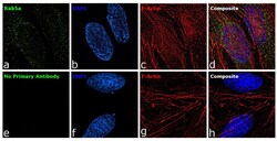
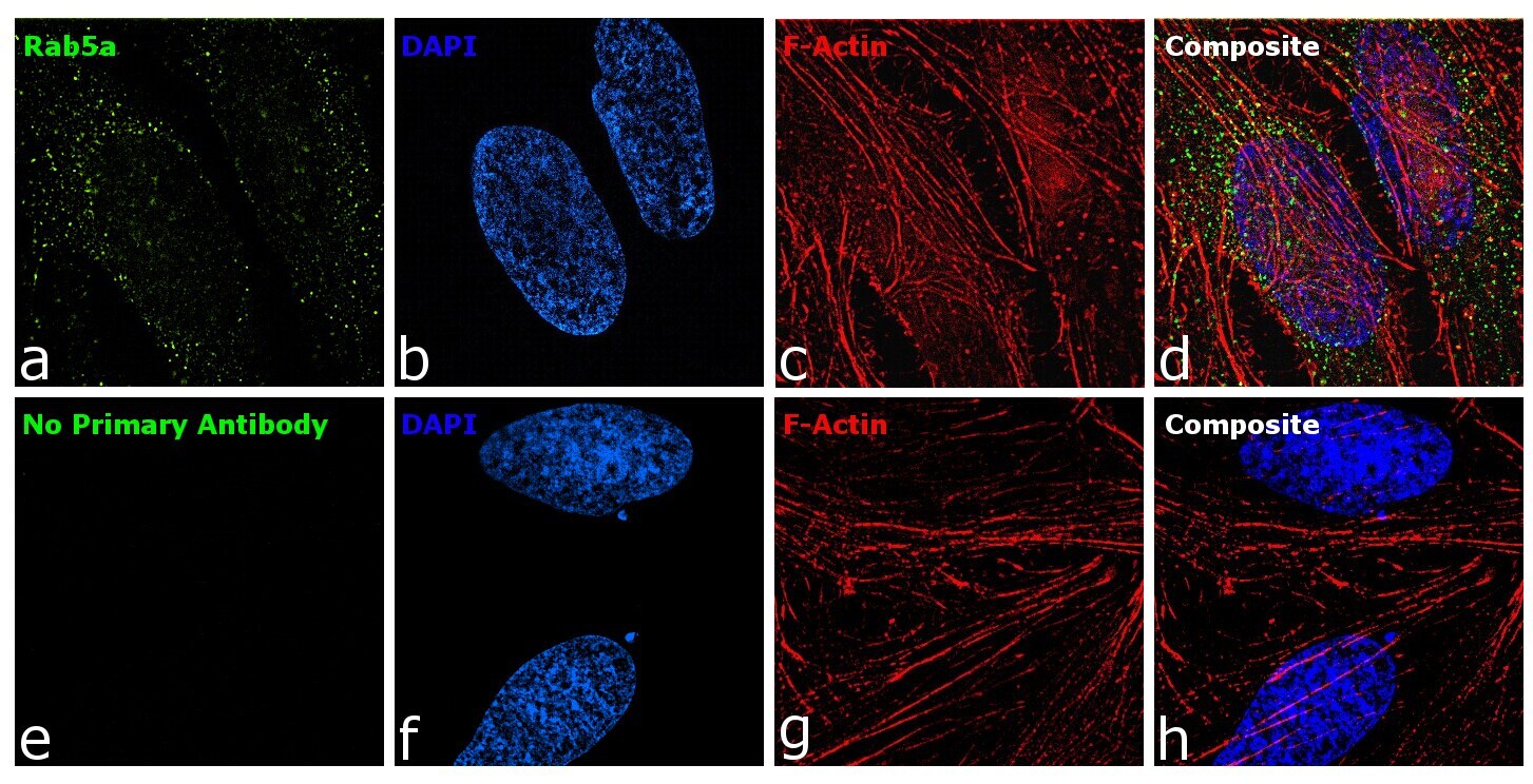
- For immunofluorescence analysis, HeLa cells were fixed for detection of endogenous Rab5a using Anti-Rab5a Monoclonal Antibody (Product # 14-9711-82, 1:100 dilution) and labeled with Donkey anti-Mouse IgG (H+L) Highly Cross-Adsorbed Secondary Antibody, Alexa Fluor Plus 488 (Product # A32766, 1:2000). Panel a) shows representative cells that were stained for detection of Rab5a protein (green), Panel b) is stained for nuclei (blue) using ProLong™ Diamond Antifade Mountant with DAPI (Product # P36962). Panel c) represents cytoskeletal F-actin staining using Rhodamine Phalloidin (Product # R415, 1:300). Panel d) is a composite image of Panels a, b and c clearly demonstrating cytoplasmic vesicular localization of Rab5a. Panel e), f), g) and h) represents control cells with no primary antibody to assess background. Images were captured at 60X magnification.
Supportive validation
- Submitted by
- Invitrogen Antibodies (provider)
- Main image
- Experimental details
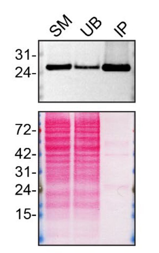
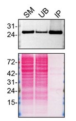
- Immunoprecipitation of RAB5A was performed on HAP1 WT cell lysate. Antibody-bead conjugate was prepared by adding 2 µg of RAB5A Monoclonal Antibody (2E8B11), eBioscience™ (Product # 14-9711-82) and 30 µL of Dynabeads™ Protein G (Product # 10004D) to 500 µl of Pierce™ IP Lysis Buffer (Product # 87788). The mixture was rocked for ~1 hour at 4 degree celcius followed by two washes to remove unbound antibodies. HAP1 WT cells were lysed in Pierce™ IP Lysis Buffer (Product # 87788) supplemented with protease inhibitor. The lysate was rocked for 30 min at 4 degree celcius and spun at 110,000xg for 15 min at 4 degree celcius. One mg of lysate was incubated with the antibody-bead conjugate for ~1 hours at 4 degree celcius. Following centrifugation and multiple washes, 4% starting material (SM), 4% unbound fraction (UB) and immunoprecipitated fraction (IP) were processed for immunoblot using a different antibody. Ponceau stained transfer of blot is shown (below immunoblot). Data courtesy of YCharOS Inc., an open science company with the mission of characterizing commercially available antibodies using knockout validation.