Explore
 Validate
 Learn
Learn Western blot
Antibody data
- Antibody Data
- Antigen structure
- References [5]
- Comments [0]
- Validations
- Western blot [1]
- Immunocytochemistry [5]
- Other assay [3]
Submit
Validation data
Reference
Comment
Report error
- Product number
- PA3-915 - Provider product page

- Provider
- Invitrogen Antibodies
- Product name
- RAB5 Polyclonal Antibody
- Antibody type
- Polyclonal
- Antigen
- Recombinant full-length protein
- Description
- PA3-915 detects Rab5 from human and rat samples. PA3-915 has been successfully used in Western blot and immunofluorescence procedures. The PA3-915 immunogen is recombinant human Rab5.
- Reactivity
- Human, Rat
- Host
- Rabbit
- Isotype
- IgG
- Vial size
- 100 µL
- Concentration
- Conc. Not Determined
- Storage
- -20° C, Avoid Freeze/Thaw Cycles
Submitted references Passive Diffusion vs Active pH-Dependent Encapsulation of Tyrosine Kinase Inhibitors Vandetanib and Lenvatinib into Folate-Targeted Ferritin Delivery System.
Efficient Protein Transfection by Swarms of Chemically Powered Plasmonic Virus-Sized Nanorobots.
Norepinephrine transporter-derived homing peptides enable rapid endocytosis of drug delivery nanovehicles into neuroblastoma cells.
Engineered mRNA-expressed antibodies prevent respiratory syncytial virus infection.
Hierarchical CRMP2 posttranslational modifications control NaV1.7 function.
Skubalova Z, Rex S, Sukupova M, Zahalka M, Skladal P, Pribyl J, Michalkova H, Weerasekera A, Adam V, Heger Z
International journal of nanomedicine 2021;16:1-14
International journal of nanomedicine 2021;16:1-14
Efficient Protein Transfection by Swarms of Chemically Powered Plasmonic Virus-Sized Nanorobots.
Ressnerova A, Novotny F, Michalkova H, Pumera M, Adam V, Heger Z
ACS nano 2021 Aug 24;15(8):12899-12910
ACS nano 2021 Aug 24;15(8):12899-12910
Norepinephrine transporter-derived homing peptides enable rapid endocytosis of drug delivery nanovehicles into neuroblastoma cells.
Haddad Y, Charousova M, Zivotska H, Splichal Z, Merlos Rodrigo MA, Michalkova H, Krizkova S, Tesarova B, Richtera L, Vitek P, Stokowa-Soltys K, Hynek D, Milosavljevic V, Rex S, Heger Z
Journal of nanobiotechnology 2020 Jul 13;18(1):95
Journal of nanobiotechnology 2020 Jul 13;18(1):95
Engineered mRNA-expressed antibodies prevent respiratory syncytial virus infection.
Tiwari PM, Vanover D, Lindsay KE, Bawage SS, Kirschman JL, Bhosle S, Lifland AW, Zurla C, Santangelo PJ
Nature communications 2018 Oct 1;9(1):3999
Nature communications 2018 Oct 1;9(1):3999
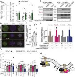
Hierarchical CRMP2 posttranslational modifications control NaV1.7 function.
Dustrude ET, Moutal A, Yang X, Wang Y, Khanna M, Khanna R
Proceedings of the National Academy of Sciences of the United States of America 2016 Dec 27;113(52):E8443-E8452
Proceedings of the National Academy of Sciences of the United States of America 2016 Dec 27;113(52):E8443-E8452
No comments: Submit comment
Supportive validation
- Submitted by
- Invitrogen Antibodies (provider)
- Main image

- Experimental details
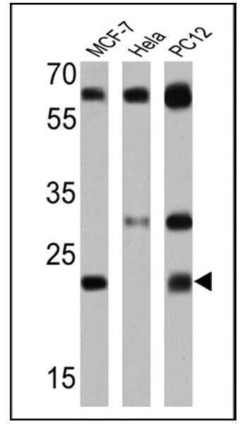
- Western blot analysis of RAB5 was performed by loading 25 µg of MCF-7 (lane 1), Hela (lane 2) and PC12 (lane 3) lysates onto an SDS polyacrylamide gel. Proteins were transferred to a PVDF membrane and blocked at 4ºC overnight. The membrane was probed with a RAB5 polyclonal antibody (Product # PA3-915) at a dilution of 1:1000 overnight at 4°C, washed in TBST, and probed with an HRP-conjugated goat anti-rabbit IgG (H+L) cross-adsorbed secondary antibody for 1 hr at room temperature in the dark. Chemiluminescent detection was performed using Pierce ECL Plus Western Blotting Substrate (Product # 32132). Results show a band at ~24 kDa in MCF-7 and PC-12 lysates.
Supportive validation
- Submitted by
- Invitrogen Antibodies (provider)
- Main image

- Experimental details
- Immunofluorescence analysis of RAB5 was done on 70% confluent log phase HeLa cells. The cells were fixed with 4% paraformaldehyde for 10 minutes, permeabilized with 0.1% Triton™ X-100 for 10 minutes, and blocked with 1% BSA for 1 hour at room temperature. The cells were labeled with RAB5 Rabbit Polyclonal Antibody (Product # PA3-915) at 1:250 dilution in 0.1% BSA and incubated for 3 hours at room temperature and then labeled with Goat anti-Rabbit IgG (H+L) Superclonal™ Secondary Antibody, Alexa Fluor® 488 conjugate (Product # A27034) at a dilution of 1:2000 for 45 minutes at room temperature (Panel a: green). Nuclei (Panel b: blue) were stained with SlowFade® Gold Antifade Mountant with DAPI (Product # S36938). F-actin (Panel c: red) was stained with Rhodamine Phalloidin (Product # R415, 1:300). Panel d is a merged image showing punctate cytoplasmic and nuclear localization. Panel e is a no primary antibody control. The images were captured at 60X magnification.

- Submitted by
- Invitrogen Antibodies (provider)
- Main image

- Experimental details
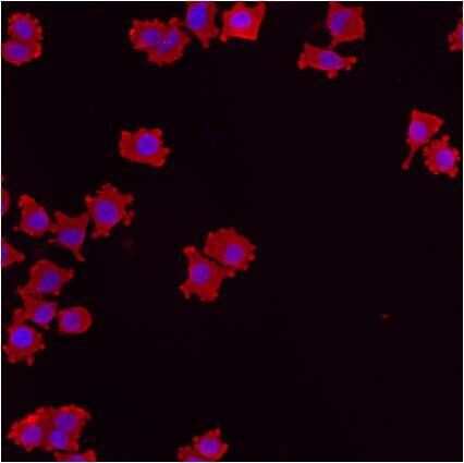
- Immunofluorescent analysis of RAB5 using anti-RAB5 polyclonal antibody (Product # PA3-915) shows staining in HMVEC Cells.

- Submitted by
- Invitrogen Antibodies (provider)
- Main image

- Experimental details
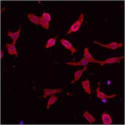
- Immunofluorescent analysis of RAB5 using anti-RAB5 polyclonal antibody (Product # PA3-915) shows staining in NS-1 Cells.
- Submitted by
- Invitrogen Antibodies (provider)
- Main image
- Experimental details
- Immunofluorescent analysis of RAB5 using anti-RAB5 polyclonal antibody (Product # PA3-915) shows staining in p19 Cells.

- Submitted by
- Invitrogen Antibodies (provider)
- Main image

- Experimental details
- Immunofluorescent analysis of RAB5 using anti-RAB5 polyclonal antibody (Product # PA3-915) shows staining in p19 Cells.

Supportive validation
- Submitted by
- Invitrogen Antibodies (provider)
- Main image
- Experimental details
- NULL

- Submitted by
- Invitrogen Antibodies (provider)
- Main image

- Experimental details
- Fig. 3 Distribution of aPali mRNAs and expression in lung epithelia. a Cy3B labeled light chain (red) and DyLight 650 labeled heavy chain (green) mRNA was transfected into the lungs of mice. At 4 h, lungs were sectioned and imaged on a whole tissue (top) or single cell basis (bottom). Scale bar represents either 200 mum (top) or 5 mum (bottom). b Tissue sections from part ( a ) were stained for Rab5 or Rab7 (green) for early or late endosomes, respectively. Scale bar represents 5 mum. Cropped regions are magnifications of white boxes in whole cell images with intensity profiles along the direction of the white arrow. c Mander's colocalization coefficient and Pearson's correlation between light chain mRNA and Rab5 or Rab7. Dotted line indicates 0 correlation. n >= 10 cells per group. Error bars represent 95% confidence interval. Asterisk indicates p < 0.05 by two-tailed t -test. d Mice lungs were transfected with either sPali (top) or aPali (bottom) mRNA using Viromer Red. At 4 h, tissue sections were stained for the expressed antibody (white). Scale bar represents 25 mum ( n = 2 mice per group). e Mice lungs were transfected with aPali heavy chain and V5 tagged light chain mRNA. Flow cytometry was performed on dissociated lungs stained for V5. Histograms of V5 intensity were normalized to the mode of intensity

- Submitted by
- Invitrogen Antibodies (provider)
- Main image

- Experimental details
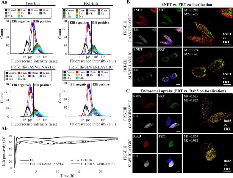
- Fig. 6 hNET-homing nanovehicles are able to induce endocytosis of hNET in SH-SY5Y cells. a Uptake kinetics of 20 uM Elli, FRT-Elli, FRT-Elli-GASNGINAYLC and FRT-Elli-SLWERLAYGIC observed over 24 h revealed by flow cytometry. Aa Elli fluorescence intensity at various time points. Ab The percentage of Elli-positive cells. b Intracellular trafficking of FRT (green) stained with Cy3 NHS ester and encapsulating Elli (80 uM, white), immunofluorescence of hNET (red) and co-localization of FRT and hNET. Hoechst 33342 was used to counterstain nuclei (blue). Scale bar, 10 um. Manders' coefficient M1 shows overlapping of hNET on FRT. Manders' coefficient M2 shows overlapping of FRT on hNET. c Co-localization of FRT (green) encapsulating Elli (white) and immunofluorescence of early endosomal marker Rab5 (red). Hoechst 33342 was used to counterstain nuclei (blue). Scale bar 10 um. Manders' coefficient M1 shows overlapping of Rab5 on FRT. Manders' coefficient M2 shows overlapping of FRT on Rab5. Manders' coefficients were taken for the representative images only