Explore
 Validate
 Learn
Learn Western blot
 Immunocytochemistry
Antibody data
- Antibody Data
- Antigen structure
- References [4]
- Comments [0]
- Validations
- Immunocytochemistry [2]
- Immunoprecipitation [1]
- Immunohistochemistry [1]
- Other assay [3]
Submit
Validation data
Reference
Comment
Report error
- Product number
- PA5-29022 - Provider product page

- Provider
- Invitrogen Antibodies
- Product name
- RAB5 Polyclonal Antibody
- Antibody type
- Polyclonal
- Antigen
- Synthetic peptide
- Description
- Recommended positive controls: A431, Raji, mouse brain. Predicted reactivity: Mouse (100%), Rat (100%), Dog (100%), Rhesus Monkey (100%). Store product as a concentrated solution. Centrifuge briefly prior to opening the vial.
- Reactivity
- Human, Mouse, Rat
- Host
- Rabbit
- Isotype
- IgG
- Vial size
- 100 μL
- Concentration
- 1.47 mg/mL
- Storage
- Store at 4°C short term. For long term storage, store at -20°C, avoiding freeze/thaw cycles.
Submitted references RAB5A expression is a predictive biomarker for trastuzumab emtansine in breast cancer.
Trappc9 deficiency in mice impairs learning and memory by causing imbalance of dopamine D1 and D2 neurons.
Cancer Alters the Metabolic Fingerprint of Extracellular Vesicles.
Cutaneous T-Cell Lymphoma (CTCL) Cell Line-Derived Extracellular Vesicles Contain HERV-W-Encoded Fusogenic Syncytin-1.
Engebraaten O, Yau C, Berg K, Borgen E, Garred Ø, Berstad MEB, Fremstedal ASV, DeMichele A, Veer LV', Esserman L, Weyergang A
Nature communications 2021 Nov 5;12(1):6427
Nature communications 2021 Nov 5;12(1):6427
Trappc9 deficiency in mice impairs learning and memory by causing imbalance of dopamine D1 and D2 neurons.
Ke Y, Weng M, Chhetri G, Usman M, Li Y, Yu Q, Ding Y, Wang Z, Wang X, Sultana P, DiFiglia M, Li X
Science advances 2020 Nov;6(47)
Science advances 2020 Nov;6(47)
Cancer Alters the Metabolic Fingerprint of Extracellular Vesicles.
Palviainen M, Laukkanen K, Tavukcuoglu Z, Velagapudi V, Kärkkäinen O, Hanhineva K, Auriola S, Ranki A, Siljander P
Cancers 2020 Nov 6;12(11)
Cancers 2020 Nov 6;12(11)
Cutaneous T-Cell Lymphoma (CTCL) Cell Line-Derived Extracellular Vesicles Contain HERV-W-Encoded Fusogenic Syncytin-1.
Laukkanen K, Saarinen M, Mallet F, Aatonen M, Hau A, Ranki A
The Journal of investigative dermatology 2020 Jul;140(7):1466-1469.e4
The Journal of investigative dermatology 2020 Jul;140(7):1466-1469.e4
No comments: Submit comment
Supportive validation
- Submitted by
- Invitrogen Antibodies (provider)
- Main image
- Experimental details
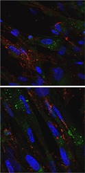
- MRC-5 cells (fetal lung fibroblasts) were grown on glass coverslips in 6 well dishes and infected for 48 h with VZV. Cells were fixed in 2% Paraformaldehyde and permeabilized with 0.02% Triton X-100 for 1 h at room temperature. Blocked cells for 2 h at RT in 5% non-fat milk plus 2.5% normal goat serum in PBS. Anti-VZV gE mouse monoclonal antibody (red) and anti-Rab5 rabbit polyclonal antibody (green, Product # PA5-29022, 1:100 in PBS) were incubated 2 h at RT (with rocking), then overnight at 4oC (with gentle rocking). After washing with PBS, cells were incubated with goat anti rabbit AlexaFluor488 (1:1250), goat anti mouse AlexaFluor546 (1:1250) and Hoechst 33342 (blue, 1:1000) for 2 h at RT (rocking, in the dark). Washed the cells with PBS. Mounted coverslips to glass and viewed on Zeiss 710 Laser Scanning Confocal Microscope. Images shown are at 400X (upper) or 630X (bottom) total magnification, respectively. Data courtesy of Dr. Grose’s lab.

- Submitted by
- Invitrogen Antibodies (provider)
- Main image

- Experimental details
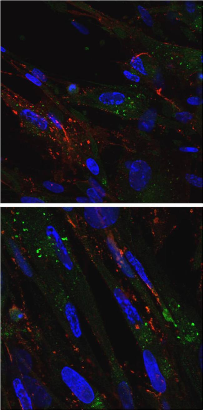
- MRC-5 cells (fetal lung fibroblasts) were grown on glass coverslips in 6 well dishes and infected for 48 h with VZV. Cells were fixed in 2% Paraformaldehyde and permeabilized with 0.02% Triton X-100 for 1 h at room temperature. Blocked cells for 2 h at RT in 5% non-fat milk plus 2.5% normal goat serum in PBS. Anti-VZV gE mouse monoclonal antibody (red) and anti-Rab5 rabbit polyclonal antibody (green, Product # PA5-29022, 1:100 in PBS) were incubated 2 h at RT (with rocking), then overnight at 4oC (with gentle rocking). After washing with PBS, cells were incubated with goat anti rabbit AlexaFluor488 (1:1250), goat anti mouse AlexaFluor546 (1:1250) and Hoechst 33342 (blue, 1:1000) for 2 h at RT (rocking, in the dark). Washed the cells with PBS. Mounted coverslips to glass and viewed on Zeiss 710 Laser Scanning Confocal Microscope. Images shown are at 400X (upper) or 630X (bottom) total magnification, respectively. Data courtesy of Dr. Groses lab.
Supportive validation
- Submitted by
- Invitrogen Antibodies (provider)
- Main image
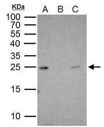
- Experimental details
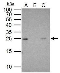
- Rab5A antibody immunoprecipitates Rab5A protein in IP experiments. IP Sample: 1 mg HeLa whole cell lysate/extract A. 30 µg HeLa whole cell lysate/extract B. Control with 2 µg of preimmune rabbit IgG C. Immunoprecipitation of Rab5A protein by 2 µg of Rab5A antibody (Product # PA5-29022) 15% SDS-PAGE The immunoprecipitated Rab5A protein was detected by Rab5A antibody (Product # PA5-29022) diluted at 1:1,000.

Supportive validation
- Submitted by
- Invitrogen Antibodies (provider)
- Main image

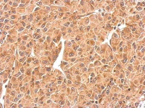
- Experimental details
- RAB5 Polyclonal Antibody detects RAB5A protein at cytosol on U87 xenograft by immunohistochemical analysis. Sample: Paraffin-embedded U87 xenograft. RAB5 Polyclonal Antibody (Product # PA5-29022) dilution: 1:500. Antigen Retrieval: EDTA based buffer, pH 8.0, 15 min.
Supportive validation
- Submitted by
- Invitrogen Antibodies (provider)
- Main image
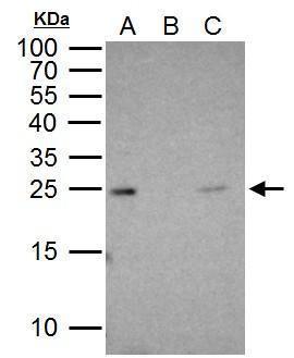
- Experimental details
- Rab5A antibody immunoprecipitates Rab5A protein in IP experiments. IP Sample: 1 mg HeLa whole cell lysate/extract A. 30 µg HeLa whole cell lysate/extract B. Control with 2 µg of preimmune rabbit IgG C. Immunoprecipitation of Rab5A protein by 2 µg of Rab5A antibody (Product # PA5-29022) 15% SDS-PAGE The immunoprecipitated Rab5A protein was detected by Rab5A antibody (Product # PA5-29022) diluted at 1:1,000.
- Submitted by
- Invitrogen Antibodies (provider)
- Main image
- Experimental details
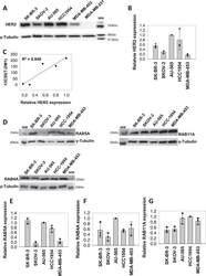
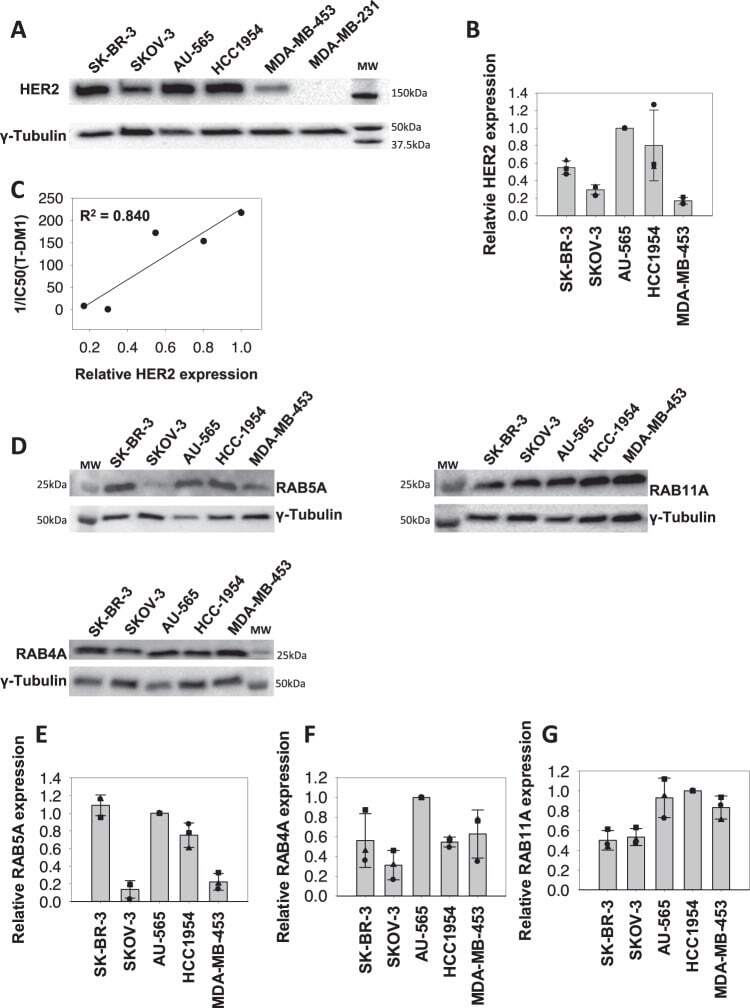
- Fig. 2 HER2 and RAB GTPase expression in cell lines. A Representative Western blot of HER2 and gamma-tubulin expression in SK-BR-3, SKOV-3, HCC1954, AU-565, MDA-MB-453, and MDA-MB-231 cells ( n = 3). B Quantification of the HER2 Western blots relative to those of gamma-tubulin. Data points represent the values of three independent experiments, the bars represent the average and the error bars represent the SD of the mean. C Linear regression analysis curve of HER2 protein expression and T-DM1 sensitivity (1/IC 50 (T-DM1)). D Representative western blot ( n = 3) of RAB4A, RAB5A, RAB11A, and gamma-tubulin expression in SK-BR-3, SKOV-3, AU-565, HCC1954, and MDA-MB-453 cells. E - G Quantification of the RAB4, RAB5, and RAB11 Western blots relative to those of gamma-tubulin. Data points represent the values of three independent experiments, the bars represent the average and the error bars represent the SD of the mean. Source data are provided as a Source Data file.
- Submitted by
- Invitrogen Antibodies (provider)
- Main image
- Experimental details
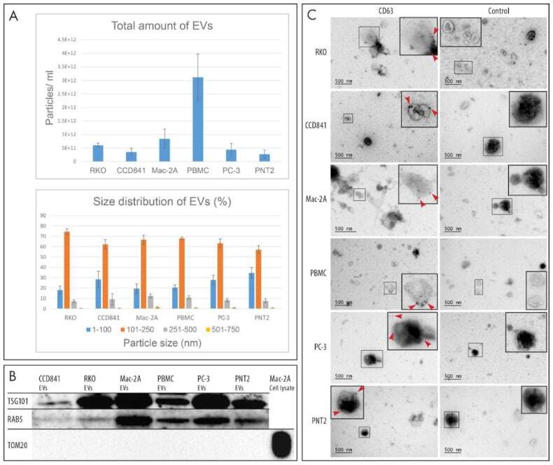
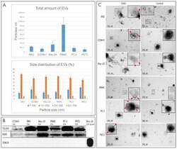
- Figure 2 Characterization of EVs. ( A ) Nanoparticle tracking analysis of total amount and size distribution of EVs. Four replicates of each EV type were analyzed. ( B ) Western blot analysis of TSG101 and RAB5 (conventional EV markers) revealed that these markers were present in all EV samples, while the mitochondrial protein TOM20 was present only in the cell lysate used as a control. Total of 1.0E10 EVs were loaded in each well. ( C ) Immunoelectron microscopy revealed the presence of the EV marker CD63 on the surface of EVs by 10-nm colloidal gold particles (red arrowheads). Immunostaining without the primary antibody was used as a negative control for CD63 immunoelectron microscopy.