Explore
 Validate
 Learn
LearnOAAB01243
antibody from Aviva Systems Biology
Targeting: KITLG
DFNA69, FPH2, Kitl, KL-1, MGF, SCF, SF, SLF
 Western blot
 ELISA
ELISAAntibody data
- Antibody Data
- Antigen structure
- References [0]
- Comments [0]
- Validations
- Western blot [2]
- Immunocytochemistry [1]
- Flow cytometry [1]
Submit
Validation data
Reference
Comment
Report error
- Product number
- OAAB01243 - Provider product page

- Provider
- Aviva Systems Biology
- Product name
- KITLG antibody - C-terminal region
- Antibody type
- Polyclonal
- Reactivity
- Human
- Host
- Rabbit
- Vial size
- 400ul
- Storage
- Maintain refrigerated at 2-8 deg C for up to 6 months. For long term storage store at -20 deg C in small aliquots to prevent freeze-thaw cycles.
No comments: Submit comment
Supportive validation
- Submitted by
- Aviva Systems Biology (provider)
- Main image
- Experimental details
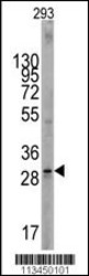
- Western blot analysis of KITLG Antibody (C-term) in 293 cell lysates (35ug/lane). KITLG (arrow) was detected using the purified Pab (1:60 dilution).
- Sample type
- 293 cell line lysates
- Primary Ab dilution
- 1.0 µg/mL
- Protocol
- Protocol

- Submitted by
- Aviva Systems Biology (provider)
- Main image
- Experimental details
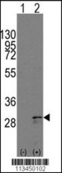
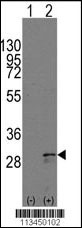
- Western blot analysis of KITLG(arrow) using rabbit polyclonal KITLG Antibody (C-term) either nontransfected (Lane 1) or lysates transiently transfected transfected with the KITLG gene (Lane 2) (Origene Technologies).
- Sample type
- lysatestransiently transfected with the KITLG gene
- Primary Ab dilution
- 1.0 µg/mL
- Protocol
- Protocol
Supportive validation
- Submitted by
- Aviva Systems Biology (provider)
- Main image

- Experimental details
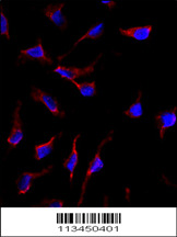
- Immunofluorescence analysis of anti-KITLG Antibody (C-term) in HeLa cells. 0.025 mg/ml primary antibody was followed by Alexa-Fluor-546-conjugated donkey anti-rabbit lgG (H+L). Alexa-Fluor-546 emits orange fluorescence. Blue counterstaining is DAPI.
- Sample type
- anti-KITLG Antibody (C-term) inHela cells. 0.025 mg/ml primary antibody was followed by Alexa-Fluor-546-conjugated donkey anti-rabbit lgG (H+L). Alexa-Fluor-546 emits orange fluorescence. Blue counterstaining is DAPI.
- Primary Ab dilution
- 1.0 µg/mL
- Protocol
- Protocol
Supportive validation
- Submitted by
- Aviva Systems Biology (provider)
- Main image

- Experimental details
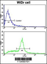
- Flow cytometric analysis of WiDr cells using SCF (KITLG) Antibody (C-term)(bottom histogram) compared to a negative control cell (top histogram). FITC-conjugated goat-anti-rabbit secondary antibodies were used for the analysis.
- Sample type
- WiDr cells
- Primary Ab dilution
- 1.0 µg/mL
- Protocol
- Protocol