Explore
 Validate
 Learn
LearnABIN2509166
antibody from antibodies-online
Targeting: KITLG
DFNA69, FPH2, Kitl, KL-1, MGF, SCF, SF, SLF
 Western blot
Antibody data
- Antibody Data
- Antigen structure
- References [0]
- Comments [0]
- Validations
- Western blot [2]
- Immunocytochemistry [1]
Submit
Validation data
Reference
Comment
Report error
- Product number
- ABIN2509166 - Provider product page

- Provider
- antibodies-online
- Product name
- anti-KIT Ligand (KITLG) antibody
- Antibody type
- Monoclonal
- Antigen
- Other
- Description
- Produced in BALB/c x ICR F1 mice using highly pure (>98%) recombinant human SCF as the immunizing antigen. This IgG1K antibody was purified from ascites fluid by Protein A affinity chromatography.
- Reactivity
- Human
- Host
- Mouse
- Vial size
- 500 μg
- Storage
- -20°C
No comments: Submit comment
Supportive validation
- Submitted by
- antibodies-online (provider)
- Main image

- Experimental details
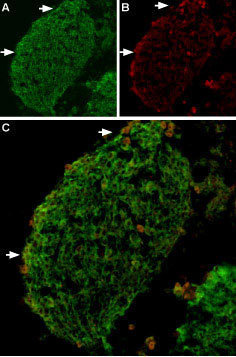
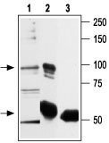
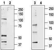
- Western blot analysis of rat brain membrane (1,2) and RBL lysates (3,4): 1,3. Anti-TRPV2 antibody (ABIN2511095), (1:200). 2,4. Anti-TRPV2 antibody, preincubated with the control peptide antigen. Immunoprecipitation of rat basophilic leukemia (RBL) lysate: 1. RBL lysate. 2. Lysate immunoprecipitated with Anti-TRPV2 antibody (ABIN2511095), (6 mg). 3. Lysate immunoprecipitated with pre-immune rabbit serum. The upper arrow indicates TRPV2 while the lower arrow indicates the IgG heavy chain. Western blot analysis was performed with Anti-TRPV2 antibody. Expression of TRPV2 in mouse DRG Immunohistochemical staining of TRPV2 in mouse dorsal root ganglion (DRG) using Anti-TRPV2 antibody (ABIN2511095). A. TRPV2 (green) appears in patches along the perimeter of the DRG (arrows). B) Neurons containing neurofilament 200 (red) are scattered in the DRG, also in patches (arrows). C. A merge of the two panels shows that the spatial distribution of neurofilament 200 and TRPV2 expression overlaps. However, DRGs showing robust expression of neurofilament 200 do not contain TRPV2.
- Submitted by
- antibodies-online (provider)
- Main image

- Experimental details
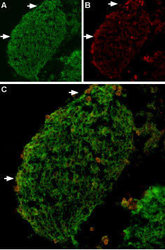
- Western blot analysis of rat brain membrane (1,2) and RBL lysates (3,4): 1,3. Anti-TRPV2 antibody (ABIN2511095), (1:200). 2,4. Anti-TRPV2 antibody, preincubated with the control peptide antigen. Immunoprecipitation of rat basophilic leukemia (RBL) lysate: 1. RBL lysate. 2. Lysate immunoprecipitated with Anti-TRPV2 antibody (ABIN2511095), (6 mg). 3. Lysate immunoprecipitated with pre-immune rabbit serum. The upper arrow indicates TRPV2 while the lower arrow indicates the IgG heavy chain. Western blot analysis was performed with Anti-TRPV2 antibody. Expression of TRPV2 in mouse DRG Immunohistochemical staining of TRPV2 in mouse dorsal root ganglion (DRG) using Anti-TRPV2 antibody (ABIN2511095). A. TRPV2 (green) appears in patches along the perimeter of the DRG (arrows). B) Neurons containing neurofilament 200 (red) are scattered in the DRG, also in patches (arrows). C. A merge of the two panels shows that the spatial distribution of neurofilament 200 and TRPV2 expression overlaps. However, DRGs showing robust expression of neurofilament 200 do not contain TRPV2.
Supportive validation
- Submitted by
- antibodies-online (provider)
- Main image
- Experimental details
- Image(s): Immunofluorescence