Explore
 Validate
 Learn
LearnPAB1884
antibody from Abnova Corporation
Targeting: KITLG
DFNA69, FPH2, Kitl, KL-1, MGF, SCF, SF, SLF
 Western blot
Antibody data
- Antibody Data
- Antigen structure
- References [3]
- Comments [0]
- Validations
- Western blot [2]
- Immunocytochemistry [1]
- Flow cytometry [1]
Submit
Validation data
Reference
Comment
Report error
- Product number
- PAB1884 - Provider product page

- Provider
- Abnova Corporation
- Proper citation
- Abnova Corporation Cat#PAB1884, RRID:AB_1575622
- Product name
- KITLG polyclonal antibody
- Antibody type
- Polyclonal
- Description
- Rabbit polyclonal antibody raised against synthetic peptide of KITLG.
- Storage
- Store at 4°C. For long term storage store at -20°C.Aliquot to avoid repeated freezing and thawing.
Submitted references Stem cell factor/c-kit receptor signaling enhances the proliferation and invasion of colorectal cancer cells through the PI3K/Akt pathway.
Differentiation of human embryonic stem cells in serum-free medium reveals distinct roles for bone morphogenetic protein 4, vascular endothelial growth factor, stem cell factor, and fibroblast growth factor 2 in hematopoiesis.
Early myeloid cells expressing c-KIT isoforms differ in signal transduction, survival and chemotactic responses to Stem Cell Factor.
Yasuda A, Sawai H, Takahashi H, Ochi N, Matsuo Y, Funahashi H, Sato M, Okada Y, Takeyama H, Manabe T
Digestive diseases and sciences 2007 Sep;52(9):2292-300
Digestive diseases and sciences 2007 Sep;52(9):2292-300
Differentiation of human embryonic stem cells in serum-free medium reveals distinct roles for bone morphogenetic protein 4, vascular endothelial growth factor, stem cell factor, and fibroblast growth factor 2 in hematopoiesis.
Pick M, Azzola L, Mossman A, Stanley EG, Elefanty AG
Stem cells (Dayton, Ohio) 2007 Sep;25(9):2206-14
Stem cells (Dayton, Ohio) 2007 Sep;25(9):2206-14
Early myeloid cells expressing c-KIT isoforms differ in signal transduction, survival and chemotactic responses to Stem Cell Factor.
Young SM, Cambareri AC, Odell A, Geary SM, Ashman LK
Cellular signalling 2007 Dec;19(12):2572-81
Cellular signalling 2007 Dec;19(12):2572-81
No comments: Submit comment
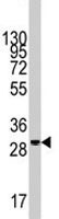
Supportive validation
- Submitted by
- Abnova Corporation (provider)
- Main image
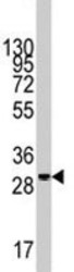
- Experimental details
- Western blot analysis of KITLG polyclonal antibody (Cat # PAB1884) in 293 cell line lysates (35 ug/lane). KITLG (arrow) was detected using the purified polyclonal antibody.
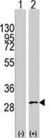
- Submitted by
- Abnova Corporation (provider)
- Main image

- Experimental details
- Western blot analysis of KITLG (arrow) using KITLG polyclonal antibody (Cat # PAB1884). 293 cell lysates (2 ug/lane) either nontransfected (Lane 1) or transiently transfected with the KITLG gene (Lane 2) (Origene Technologies).
Supportive validation
- Submitted by
- Abnova Corporation (provider)
- Main image
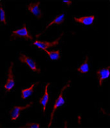
- Experimental details
- Immunofluorescence analysis of KITLG polyclonal antibody (Cat # PAB1884) in HeLa cells. 0.025 mg/mL primary antibody was followed by Alexa-Fluor-546-conjugated donkey anti-rabbit lgG (H+L). Alexa-Fluor-546 emits orange fluorescence. Blue counterstaining is DAPI.
- Validation comment
- Immunofluorescence

Supportive validation
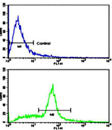
- Submitted by
- Abnova Corporation (provider)
- Main image

- Experimental details
- Flow cytometric analysis of WiDr cells using KITLG polyclonal antibody (Cat # PAB1884)(bottom histogram) compared to a negative control cell (top histogram).FITC-conjugated goat-anti-rabbit secondary antibodies were used for the analysis.
- Validation comment
- Flow Cytometry