Explore
 Validate
 Learn
Learn Flow cytometry
Antibody data
- Antibody Data
- Antigen structure
- References [2]
- Comments [0]
- Validations
- Flow cytometry [1]
Submit
Validation data
Reference
Comment
Report error
- Product number
- 17-9037-41 - Provider product page

- Provider
- Invitrogen Antibodies
- Product name
- Anti-Phospho-SLP-76 (Tyr128) Monoclonal Antibody (HNDZ55), APC, eBioscience™
- Antibody type
- Monoclonal
- Antigen
- Other
- Description
- Description: This HNDZ55 monoclonal antibody recognizes human SH2 domain-containing leukocyte protein of 76 kD (SLP-76) when phosphorylated on tyrosine 128 (Y128). SLP-76 is phosphorylated by ZAP-70 downstream of the T cell receptor (TCR). SLP-76 contains an acidic region that includes a PEST domain, several tyrosine residues that are phosphorylated following TCR ligation, a proline-rich domain, and an SH2 domain. Numerous proteins associate with SLP-76 including Vav1, GADS, and PLC gamma 1, supporting the notion that SLP-76 functions as a critical adaptor protein downstream of the TCR. SLP-76-deficient T cell lines or mice deficient in SLP-76 have shown that SLP-76 plays a positive role in promoting T cell development and activation as well as mast cell and platelet function. This antibody recognizes only human SLP-76 when phosphorylated on Y128. It is not recommended for staining of mouse cells. Applications Reported: This HNDZ55 antibody has been reported for use in intracellular staining followed by flow cytometric analysis. Applications Tested: This HNDZ55 antibody has been pre-titrated and tested by intracellular staining followed by flow cytometric analysis of stimulated normal human peripheral blood cells. This can be used at 5 µL (0.5 µg) per test. A test is defined as the amount (µg) of antibody that will stain a cell sample in a final volume of 100 µL. Cell number should be determined empirically but can range from 10^5 to 10^8 cells/test. Staining Protocol: Protocol A and Protocol C are recommended for this monoclonal antibody. Use of Protocol A: Two-step protocol: intracellular (cytoplasmic) proteins allows for the greatest flexibility for detection of surface and intracellular (cytoplasmic) proteins. Protocol C: Two-step protocol: Fixation/Methanol allows for the greatest discrimination of phospho-specific signaling between unstimulated and stimulated samples, but with limitations on the ability to stain specific surface proteins (refer to "Clone Performance Following Fixation/Permeabilization" located in the Best Protocols Section under the Resources tab online). All Protocols can be found in the Flow Cytometry Protocols: "Staining Intracellular Antigens for Flow Cytometry Protocol" located in the Best Protocols Section under the Resources tab online. Excitation: 633-647 nm; Emission: 660 nm; Laser: Red Laser. Filtration: 0.2 µm post-manufacturing filtered.
- Reactivity
- Human
- Host
- Mouse
- Isotype
- IgG
- Antibody clone number
- HNDZ55
- Vial size
- 25 Tests
- Concentration
- 5 µL/Test
- Storage
- 4° C, store in dark, DO NOT FREEZE!
Submitted references The SLP-76 Src homology 2 domain is required for T cell development and activation.
Conditional deletion of SLP-76 in mature T cells abrogates peripheral immune responses.
Burns JC, Corbo E, Degen J, Gohil M, Anterasian C, Schraven B, Koretzky GA, Kliche S, Jordan MS
Journal of immunology (Baltimore, Md. : 1950) 2011 Nov 1;187(9):4459-66
Journal of immunology (Baltimore, Md. : 1950) 2011 Nov 1;187(9):4459-66
Conditional deletion of SLP-76 in mature T cells abrogates peripheral immune responses.
Wu GF, Corbo E, Schmidt M, Smith-Garvin JE, Riese MJ, Jordan MS, Laufer TM, Brown EJ, Maltzman JS
European journal of immunology 2011 Jul;41(7):2064-73
European journal of immunology 2011 Jul;41(7):2064-73
No comments: Submit comment
Supportive validation
- Submitted by
- Invitrogen Antibodies (provider)
- Main image
- Experimental details
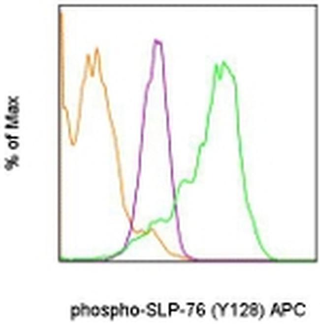
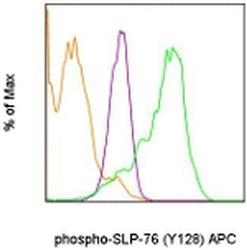
- Intracellular staining of normal human peripheral blood cells that were unstimulated (orange histogram), stimulated with Anti-Human CD3 Functional Grade Purified (Product # 16-0037-81) and goat anti-mouse IgG for 5 minutes (purple histogram), or treated with hydrogen peroxide-activated sodium pervanadate (green histogram) with Anti-Human phospho-SLP-76 (Y128) APC. The Intracellular Fixation & Permeabilization Buffer Set (88-8824) and protocol were used for staining. Lymphocytes in the CD3+ gate were used for analysis.