Explore
 Validate
 Learn
Learn Western blot
 Immunohistochemistry
Antibody data
- Antibody Data
- Antigen structure
- References [0]
- Comments [0]
- Validations
- Western blot [2]
- Immunocytochemistry [2]
Submit
Validation data
Reference
Comment
Report error
- Product number
- MA1-19369 - Provider product page

- Provider
- Invitrogen Antibodies
- Product name
- SLP76 Monoclonal Antibody (SLP-76/03)
- Antibody type
- Monoclonal
- Antigen
- Other
- Description
- This antibody reacts with SLP76, a 76kDa cytosolic adaptor protein that is involved in signaling of various hematopoietic cells, such as T cells, mast cells or neutrophils; in B cells, however, it is replaced by SLP65.
- Reactivity
- Human, Mouse, Porcine
- Host
- Mouse
- Isotype
- IgG
- Antibody clone number
- SLP-76/03
- Vial size
- 100 μg
- Concentration
- 1 mg/mL
- Storage
- 4°C, do not freeze
No comments: Submit comment
Supportive validation
- Submitted by
- Invitrogen Antibodies (provider)
- Main image

- Experimental details
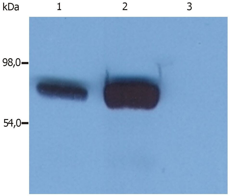
- Western blotting analysis (reducing conditions) of whole cell lysate using anti-SLP76 (SLP-76/03) Monoclonal antibody (Product # MA1-19369). Lane 1: HPB-ALL human peripheral blood T cell leukemia cell line; Lane 2: JURKAT human peripheral blood T cell leukemia cell line; Lane 3: RAMOS human Burkitt lymphoma cell line.
- Submitted by
- Invitrogen Antibodies (provider)
- Main image

- Experimental details
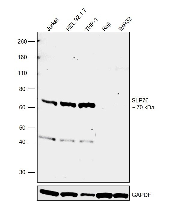
- Western blot was performed using Anti-SLP76 Monoclonal Antibody (SLP-76/03) (Product # MA1-19369) and a 70 kDa band corresponding to SLP76 was observed across the cell lines tested except in Raji and IMR-32. Whole cell extracts (30 µg lysate) of Jurkat (Lane 1), HEL 92.1.7 (Lane 2), THP-1 (Lane 3), Raji (Lane 4), and IMR32 (Lane 5) were electrophoresed using NuPAGE™ 4-12% Bis-Tris Protein Gel (Product # NP0322BOX). Resolved proteins were then transferred onto a nitrocellulose membrane (Product # IB23001) by iBlot® 2 Dry Blotting System (Product # IB21001). The blot was probed with the primary antibody (1:1000 dilution) and detected by chemiluminescence with Goat anti-Mouse IgG (H+L) Superclonal™ Recombinant Secondary Antibody, HRP (Product # A28177, 1:4000 dilution) using the iBright FL 1000 (Product # A32752). Chemiluminescent detection was performed using Novex® ECL Chemiluminescent Substrate Reagent Kit (Product # WP20005).
Supportive validation
- Submitted by
- Invitrogen Antibodies (provider)
- Main image

- Experimental details
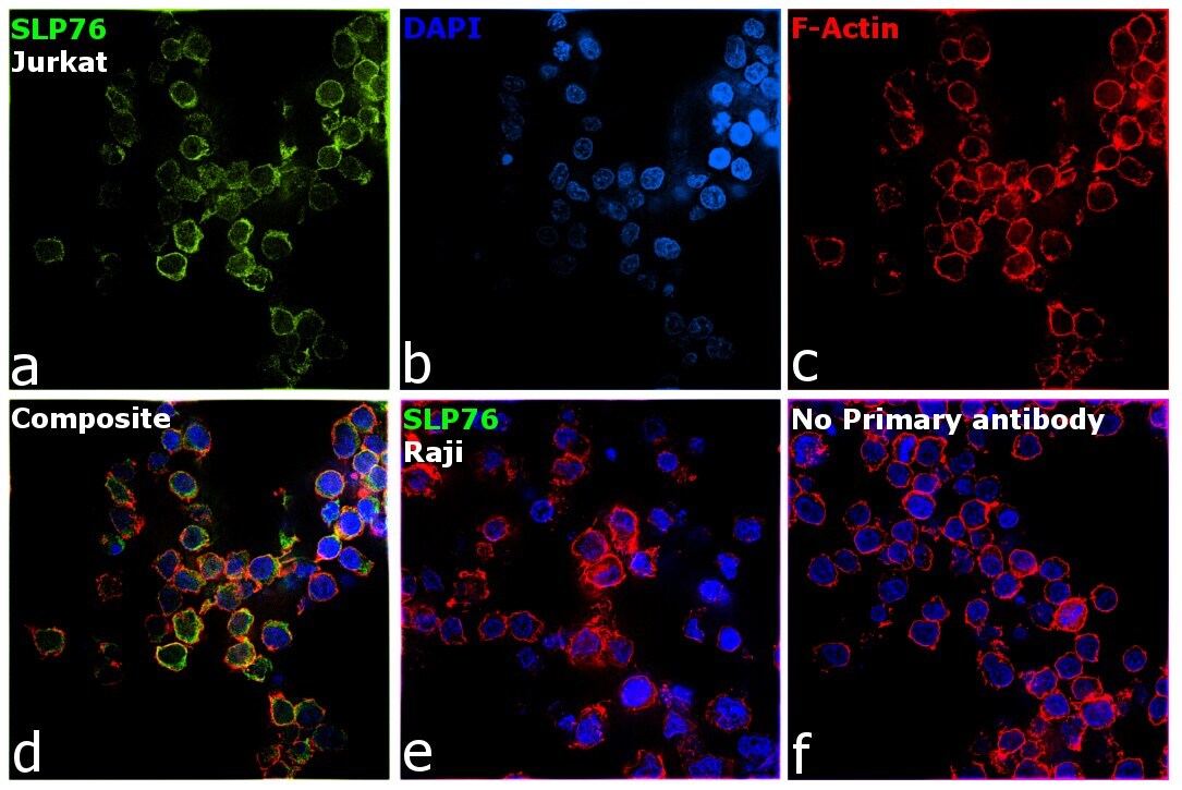
- Immunofluorescence analysis of SLP76 was performed using 70% confluent log phase Jurkat cells. The cells were fixed with 4% paraformaldehyde for 10 minutes, permeabilized with 0.1% Triton™ X-100 for 15 minutes, and blocked with 2% BSA for 1 hour at room temperature. The cells were labeled with SLP76 Monoclonal Antibody (SLP-76/03) (Product # MA1-19369) at 1:100 dilution in 0.1% BSA, incubated at 4 degree Celsius overnight and then with Donkey anti-Mouse IgG (H+L) Highly Cross-Adsorbed Secondary Antibody, Alexa Fluor Plus 488 (Product # A32766) at a dilution of 1:2000 for 45 minutes at room temperature (Panel a: green). Nuclei (Panel b: blue) were stained with SlowFade® Gold Antifade Mountant with DAPI (Product # S36938). F-actin (Panel c: red) was stained with Rhodamine Phalloidin (Product # R415, 1:300). Panel d represents the merged image showing cytoplasmic localization. Panel e represents Raji cells having no expression of SLP76. Panel f represents control cells with no primary antibody to assess background. The images were captured at 60X magnification.

- Submitted by
- Invitrogen Antibodies (provider)
- Main image

- Experimental details
- Immunofluorescence analysis of SLP76 was performed using 70% confluent log phase Jurkat cells. The cells were fixed with 4% paraformaldehyde for 10 minutes, permeabilized with 0.1% Triton™ X-100 for 15 minutes, and blocked with 2% BSA for 1 hour at room temperature. The cells were labeled with SLP76 Monoclonal Antibody (SLP-76/03) (Product # MA1-19369) at 1:100 dilution in 0.1% BSA, incubated at 4 degree Celsius overnight and then with Donkey anti-Mouse IgG (H+L) Highly Cross-Adsorbed Secondary Antibody, Alexa Fluor Plus 488 (Product # A32766) at a dilution of 1:2000 for 45 minutes at room temperature (Panel a: green). Nuclei (Panel b: blue) were stained with SlowFade® Gold Antifade Mountant with DAPI (Product # S36938). F-actin (Panel c: red) was stained with Rhodamine Phalloidin (Product # R415, 1:300). Panel d represents the merged image showing cytoplasmic localization. Panel e represents Raji cells having no expression of SLP76. Panel f represents control cells with no primary antibody to assess background. The images were captured at 60X magnification.