 Explore
 Validate
 Learn
Learn Western blot
Antibody data
- Antibody Data
- Antigen structure
- References [0]
- Comments [0]
- Validations
- Western blot [4]
- Immunocytochemistry [1]
- Immunoprecipitation [1]
- Immunohistochemistry [2]
Submit
Validation data
Reference
Comment
Report error
- Product number
- GTX129426 - Provider product page

- Provider
- GeneTex
- Product name
- SP3 antibody
- Antibody type
- Polyclonal
- Reactivity
- Human
- Host
- Rabbit
No comments: Submit comment
Supportive validation
- Submitted by
- GeneTex (provider)
- Main image
- Experimental details
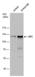
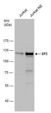
- SP3 antibody detects SP3 protein by western blot analysis.A. 30 £gg Jurkat whole cell extractB. 30 £gg Jurkat nuclear extract7.5 % SDS-PAGESP3 antibody (GTX129426) dilution: 1:1000
- Submitted by
- GeneTex (provider)
- Main image
- Experimental details
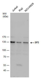
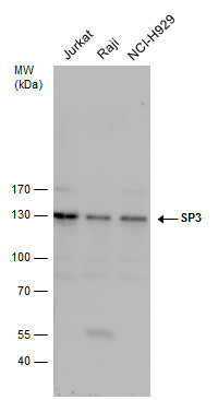
- SP3 antibody detects SP3 protein by western blot analysis. Various whole cell extracts (30 £gg) were separated by 7.5 % SDS-PAGE, and blotted with SP3 antibody (GTX129426) diluted by 1:1000
- Submitted by
- GeneTex (provider)
- Main image
- Experimental details
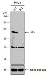
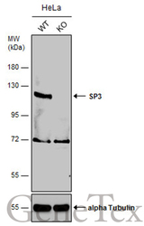
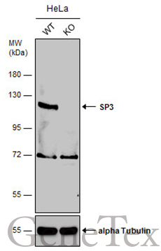
- Wild-type (WT) and SP3 knockout (KO) HeLa cell extracts (30 ?g) were separated by 7.5% SDS-PAGE, and the membrane was blotted with SP3 antibody (GTX129426) diluted at 1:1000. The HRP-conjugated anti-rabbit IgG antibody (GTX213110-01) was used to detect the primary antibody, and the signal was developed with Trident ECL plus-Enhanced.
- Submitted by
- GeneTex (provider)
- Main image
- Experimental details
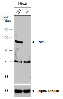
- Wild-type (WT) and SP3 knockout (KO) HeLa cell extracts (30 ?g) were separated by 7.5% SDS-PAGE, and the membrane was blotted with SP3 antibody (GTX129426) diluted at 1:1000. The HRP-conjugated anti-rabbit IgG antibody (GTX213110-01) was used to detect the primary antibody, and the signal was developed with Trident ECL plus-Enhanced.
Supportive validation
- Submitted by
- GeneTex (provider)
- Main image
- Experimental details
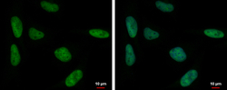
- SP3 antibody detects SP3 protein at nucleus by immunofluorescent analysis.Sample: HeLa cells were fixed in 4% paraformaldehyde at RT for 5 min.Green: SP3 protein stained by SP3 antibody (GTX129426) diluted at 1:500.Blue: Hoechst 33342 staining.

Supportive validation
- Submitted by
- GeneTex (provider)
- Main image
- Experimental details
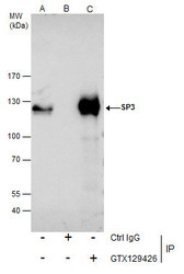
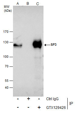
- Immunoprecipitation of SP3 protein from Jurkat nuclear extracts using 5 £gg of SP3 antibody (GTX129426).Western blot analysis was performed using SP3 antibody (GTX129426).EasyBlot anti-Rabbit IgG (GTX221666-01) was used as a secondary reagent.
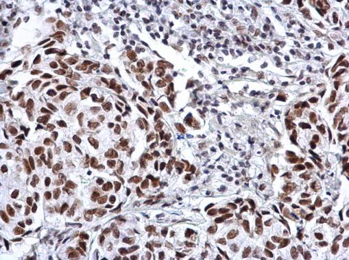
Supportive validation
- Submitted by
- GeneTex (provider)
- Main image
- Experimental details
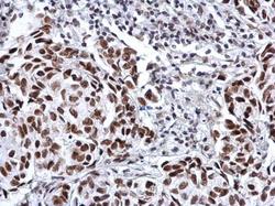
- SP3 antibody detects SP3 protein at nucleus on human breast carcinoma by immunohistochemical analysis. Sample: Paraffin-embedded human breast carcinoma. SP3 antibody (GTX129426) dilution: 1:1000.
- Submitted by
- GeneTex (provider)
- Main image
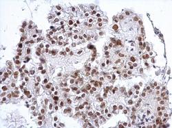
- Experimental details
- SP3 antibody detects SP3 protein at nucleus on human ovarian carcinoma by immunohistochemical analysis. Sample: Paraffin-embedded human ovarian carcinoma. SP3 antibody (GTX129426) dilution: 1:1000.
