Explore
 Validate
 Learn
Learn Western blot
 ELISA
ELISA Immunocytochemistry
Antibody data
- Antibody Data
- Antigen structure
- References [0]
- Comments [0]
- Validations
- Immunocytochemistry [2]
Submit
Validation data
Reference
Comment
Report error
- Product number
- MA5-20003 - Provider product page

- Provider
- Invitrogen Antibodies
- Product name
- SP3 Monoclonal Antibody (4E5)
- Antibody type
- Monoclonal
- Antigen
- Recombinant full-length protein
- Description
- Peptide Sequence: TAGINADGHL INTGQAMDSS DNSERTGERV SPDINETNTD TDLFVPTSSS SQLPVTIDST GILQQNTNSL TTSSGQVHSS DLQGNYIQSP VSEETQAQNI QVSTAQPVVQ HLQLQESQQP TSQAQIVQGI TPQTIHGVQA SGQN*
- Reactivity
- Human, Mouse, Rat
- Host
- Mouse
- Isotype
- IgG
- Antibody clone number
- 4E5
- Vial size
- 100 μg
- Concentration
- 1 mg/mL
- Storage
- -20°C, Avoid Freeze/Thaw Cycles
No comments: Submit comment
Supportive validation
- Submitted by
- Invitrogen Antibodies (provider)
- Main image
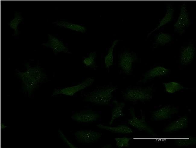
- Experimental details
- Immunofluorescence analysis of HeLa cells using an anti-SP3 monoclonal antibody (Product # MA5-20003). (antibody concentration 10 µg/mL).
- Submitted by
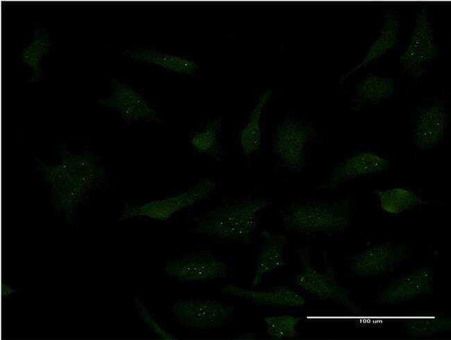
- Invitrogen Antibodies (provider)
- Main image
- Experimental details
- Immunofluorescence analysis of HeLa cells using an anti-SP3 monoclonal antibody (Product # MA5-20003). (antibody concentration 10 µg/mL).