 Explore
 Validate
 Learn
Learn Western blot
 Immunocytochemistry
Antibody data
- Antibody Data
- Antigen structure
- References [1]
- Comments [0]
- Validations
- Immunocytochemistry [3]
- Immunohistochemistry [3]
Submit
Validation data
Reference
Comment
Report error
- Product number
- PA5-78176 - Provider product page

- Provider
- Invitrogen Antibodies
- Product name
- SP3 Polyclonal Antibody
- Antibody type
- Polyclonal
- Antigen
- Recombinant full-length protein
- Description
- Positive Control: HeLa, Jurkat, Jurkat_NE, Raji, NCI-H929 Store product as a concentrated solution. Centrifuge briefly prior to opening the vial.
- Reactivity
- Human
- Host
- Rabbit
- Isotype
- IgG
- Vial size
- 100 μL
- Concentration
- 0.2 mg/mL
- Storage
- Store at 4°C short term. For long term storage, store at -20°C, avoiding freeze/thaw cycles.
Submitted references A novel Cbx1, PurB, and Sp3 complex mediates long-term silencing of tissue- and lineage-specific genes.
Baksh SS, Pratt RE, Gomez J, Dzau VJ, Hodgkinson CP
The Journal of biological chemistry 2022 Jun;298(6):102053
The Journal of biological chemistry 2022 Jun;298(6):102053
No comments: Submit comment
Supportive validation
- Submitted by
- Invitrogen Antibodies (provider)
- Main image

- Experimental details
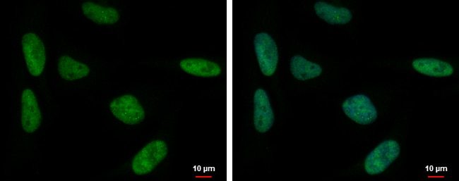
- Immunofluorescent analysis of SP3 in HeLa cells. Samples were treated with 4% paraformaldehyde at RT for 5 min and incubated with SP3 polyclonal antibody (Product # PA5-78176) using a dilution of 1:500, followed by Hoechst.
- Submitted by
- Invitrogen Antibodies (provider)
- Main image

- Experimental details
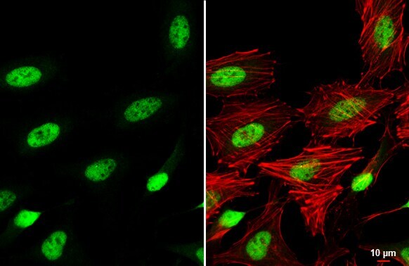
- SP3 Polyclonal Antibody detects SP3 protein at nucleus by immunofluorescent analysis. Sample: HeLa cells were fixed in 4% paraformaldehyde at RT for 15 min. Green: SP3 stained by SP3 Polyclonal Antibody (Product # PA5-78176) diluted at 1:500. Red: phalloidin, a cytoskeleton marker, diluted at 1:200. Scale bar= 10 µm.

- Submitted by
- Invitrogen Antibodies (provider)
- Main image

- Experimental details
- SP3 Polyclonal Antibody detects SP3 protein at nucleus by immunofluorescent analysis. Sample: HeLa cells were fixed in 4% paraformaldehyde at RT for 15 min. Green: SP3 stained by SP3 Polyclonal Antibody (Product # PA5-78176) diluted at 1:500. Red: phalloidin, a cytoskeleton marker, diluted at 1:200. Scale bar= 10 µm.
Supportive validation
- Submitted by
- Invitrogen Antibodies (provider)
- Main image
- Experimental details
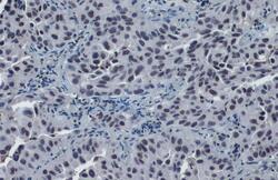
- SP3 Polyclonal Antibody detects SP3 protein at nucleus by immunohistochemical analysis. Sample: Paraffin-embedded human lung cancer. SP3 stained by SP3 Polyclonal Antibody (Product # PA5-78176) diluted at 1:500. Antigen Retrieval: Citrate buffer, pH 6.0, 15 min.
- Submitted by
- Invitrogen Antibodies (provider)
- Main image

- Experimental details
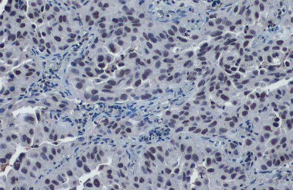
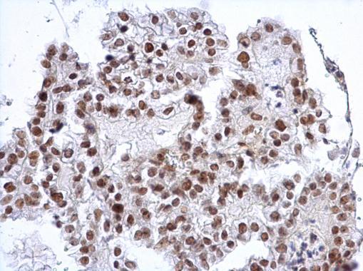
- SP3 Polyclonal Antibody detects SP3 protein at nucleus on human ovarian carcinoma by immunohistochemical analysis. Sample: Paraffin-embedded human ovarian carcinoma. SP3 Polyclonal Antibody (Product # PA5-78176) dilution: 1:1,000. Antigen Retrieval: EDTA based buffer, pH 8.0, 15 min.
- Submitted by
- Invitrogen Antibodies (provider)
- Main image
- Experimental details
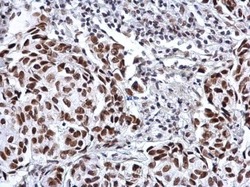
- SP3 Polyclonal Antibody detects SP3 protein at nucleus on human breast carcinoma by immunohistochemical analysis. Sample: Paraffin-embedded human breast carcinoma. SP3 Polyclonal Antibody (Product # PA5-78176) dilution: 1:1,000. Antigen Retrieval: EDTA based buffer, pH 8.0, 15 min.
