Explore
 Validate
 Learn
Learn Western blot
Antibody data
- Antibody Data
- Antigen structure
- References [2]
- Comments [0]
- Validations
- Western blot [4]
- Immunocytochemistry [3]
- Other assay [1]
Submit
Validation data
Reference
Comment
Report error
- Product number
- PA1-28042 - Provider product page

- Provider
- Invitrogen Antibodies
- Product name
- gamma Tubulin Polyclonal Antibody
- Antibody type
- Polyclonal
- Antigen
- Synthetic peptide
- Description
- Recommended positive controls: chicken fibroblast, A431.
- Concentration
- 10.5 mg/mL
Submitted references Induced Arp2/3 Complex Depletion Increases FMNL2/3 Formin Expression and Filopodia Formation.
A homozygous deleterious CDK10 mutation in a patient with agenesis of corpus callosum, retinopathy, and deafness.
Dimchev V, Lahmann I, Koestler SA, Kage F, Dimchev G, Steffen A, Stradal TEB, Vauti F, Arnold HH, Rottner K
Frontiers in cell and developmental biology 2021;9:634708
Frontiers in cell and developmental biology 2021;9:634708
A homozygous deleterious CDK10 mutation in a patient with agenesis of corpus callosum, retinopathy, and deafness.
Guen VJ, Edvardson S, Fraenkel ND, Fattal-Valevski A, Jalas C, Anteby I, Shaag A, Dor T, Gillis D, Kerem E, Lees JA, Colas P, Elpeleg O
American journal of medical genetics. Part A 2018 Jan;176(1):92-98
American journal of medical genetics. Part A 2018 Jan;176(1):92-98
No comments: Submit comment
Supportive validation
- Submitted by
- Invitrogen Antibodies (provider)
- Main image
- Experimental details
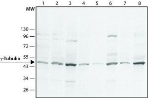
- Western Blot analysis of gamma Tubulin was performed by loading (1) A431 (2) HeLa (3) Jurkat (4) C2C12 (5) rat2 (6) U87 (7) NIH3T3 (8) CHO cells. Proteins were transferred to a membrane and probed with a gamma Tubulin Polyclonal Antibody (Product # PA1-28042) at a dilution of 1:1,000.

- Submitted by
- Invitrogen Antibodies (provider)
- Main image

- Experimental details
- Western Blot analysis of gamma Tubulin was performed by loading (1) A431 (2) HeLa (3) Jurkat (4) C2C12 (5) rat2 (6) U87 (7) NIH3T3 (8) CHO cells. Proteins were transferred to a membrane and probed with a gamma Tubulin Polyclonal Antibody (Product # PA1-28042) at a dilution of 1:1,000.
- Submitted by
- Invitrogen Antibodies (provider)
- Main image

- Experimental details
- Knockdown of gamma Tubulin was achieved by transfecting HeLa cells with gamma Tubulin specific siRNAs (Silencer® select Product # s14502). Western blot analysis (Fig. a) was performed using membrane extracts from the gamma Tubulin knockdown cells (lane 3), non-specific scrambled siRNA transfected cells (lane 2) and untransfected cells (lane 1). The blots were probed with gamma Tubulin Polyclonal Antibody (Product # PA1-28042, 1:1000 dilution) and Goat anti-Rabbit IgG (H+L) Superclonal™ Secondary Antibody, HRP conjugate (Product # A27036, 0.25 µg/mL, 1:4000 dilution). Densitometric analysis of this western blot is shown in histogram (Fig. b). Decrease in signal upon siRNA mediated knock down confirms that antibody is specific to gamma Tubulin.

- Submitted by
- Invitrogen Antibodies (provider)
- Main image

- Experimental details
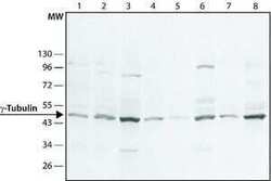
- Western blot analysis was performed on whole cell extracts (30 µg lysate) of HeLa (Lane 1), MCF7 (Lane 2), A549 (Lane 3), Hep G2 (Lane 4), Caco-2 (Lane 5), A-431 (Lane 6), C2C12 (Lane 7), NIH/3T3 (Lane 8) and SH-SY5Y (Lane 9). The blot was probed with Anti-gamma Tubulin Polyclonal Antibody (Product # PA1-28042, 1:1000 dilution) and detected by chemiluminescence using Goat anti Rabbit IgG (H+L) Superclonal™ Secondary Antibody, HRP conjugate (Product # A27036, 0.25 µg/mL, 1:4000 dilution). A 51 kDa band corresponding to gamma Tubulin was observed across all cell lines tested.

Supportive validation
- Submitted by
- Invitrogen Antibodies (provider)
- Main image

- Experimental details
- Immunocytochemistry-Immunofluorescence analysis of gamma Tubulin in NIH3T3 cells using gamma Tubulin Polyclonal Antibody (Product # PA1 28042) at 1:500 (green). Actin staining with phalloidin rhodamine conjugate (red). Nuclear staining with DAPI (blue).

- Submitted by
- Invitrogen Antibodies (provider)
- Main image
- Experimental details
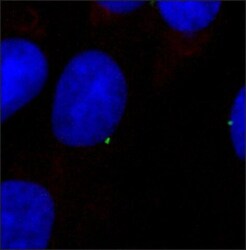
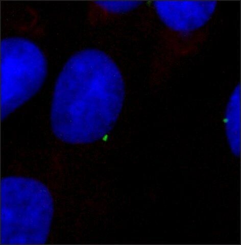
- Immunocytochemistry-Immunofluorescence analysis of gamma Tubulin in Rat2 cells using gamma Tubulin Polyclonal Antibody (Product # PA1 28042) at 1:1500 (green) with DAPI (blue).
- Submitted by
- Invitrogen Antibodies (provider)
- Main image

- Experimental details
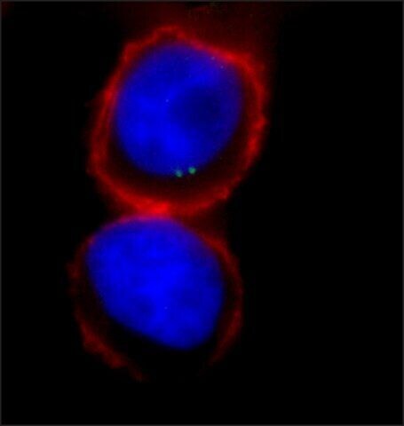
- Immunocytochemistry-Immunofluorescence analysis of gamma Tubulin in HeLa cells using gamma Tubulin Polyclonal Antibody (Product # PA1 28042) at 1:500(green). Actin staining with phalloidin rhodamine conjugate (red). Nuclear staining with DAPI (blue).
Supportive validation
- Submitted by
- Invitrogen Antibodies (provider)
- Main image
- Experimental details
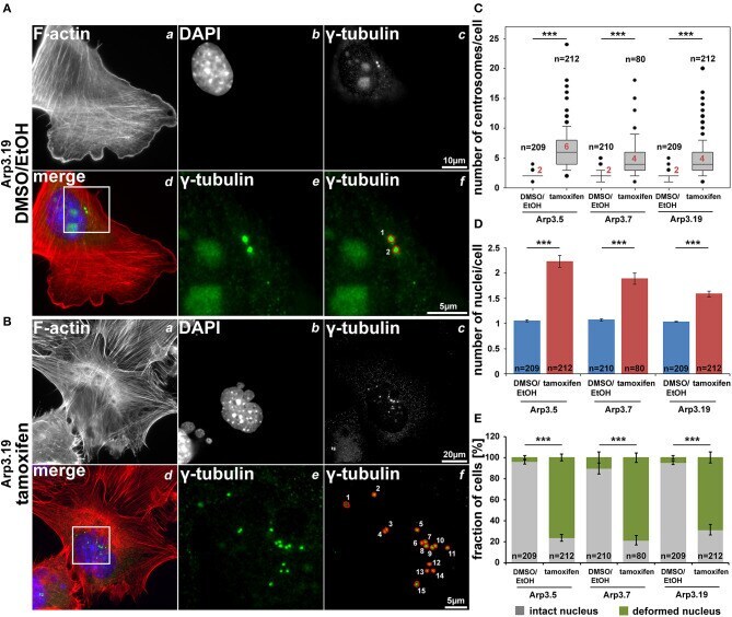
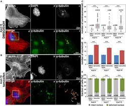
- Figure 10 Arp3 depletion causes an increase of centrosome and nucleus numbers as well as nuclear deformation. (A,B) Representative fluorescence microscopy images of Actr3 fl / fl cells (clone Arp3.19) stained for the F-actin cytoskeleton with phalloidin (a) , nuclei using DAPI (b) and gamma-tubulin for centrosomes (c) . Merged images (d) display actin filaments in red, nuclei in blue and centrosomes in green. White rectangles in (d) mark regions of interest enlarged in (e) displaying centrosomes, the quantification of which is illustrated in (f) . (C,D) Box and whiskers plots (as described for Figure 4B ) displaying numbers of centrosomes (C) or of nuclei (D) . For statistics, non-parametric, Mann-Whitney rank sum tests were used (*** p < 0.001). (E) Analysis of nuclear morphologies. Cells were categorized according to the morphological integrity of their nuclei (intact nuclei in gray; deformed nuclei in green), represented as a fraction of all cells analyzed in individual cell populations. Results are depicted as stacked columns representing arithmetic means and SEMs from three independent experiments; non-parametric, Mann-Whitney rank sum test for statistics (*** p < 0.001), n = total number of cells analyzed in (C-E) .