 Explore
 Validate
 Learn
Learn Western blot
 Immunocytochemistry
Antibody data
- Antibody Data
- Antigen structure
- References [0]
- Comments [0]
- Validations
- Immunocytochemistry [4]
- Immunoprecipitation [1]
- Immunohistochemistry [1]
- Other assay [2]
Submit
Validation data
Reference
Comment
Report error
- Product number
- PA5-34815 - Provider product page

- Provider
- Invitrogen Antibodies
- Product name
- gamma Tubulin Polyclonal Antibody
- Antibody type
- Polyclonal
- Antigen
- Recombinant full-length protein
- Description
- Recommended positive controls: 293T, A431, HeLa, Jurkat, Raji, NCI-H929, NIH3T3, rat kidney. Predicted reactivity: Mouse (99%), Rat (99%), Zebrafish (98%), Xenopus laevis (99%), Dog (100%), Rhesus Monkey (100%), Chimpanzee (100%), Bovine (100%). Store product as a concentrated solution. Centrifuge briefly prior to opening the vial.
- Reactivity
- Human, Mouse, Rat, Zebrafish
- Host
- Rabbit
- Isotype
- IgG
- Vial size
- 100 μL
- Concentration
- 0.24 mg/mL
- Storage
- Store at 4°C short term. For long term storage, store at -20°C, avoiding freeze/thaw cycles.
No comments: Submit comment
Supportive validation
- Submitted by
- Invitrogen Antibodies (provider)
- Main image
- Experimental details
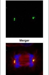
- Immunofluorescent analysis of gamma Tubulin in methanol-fixed 293T cells using a gamma Tubulin polyclonal antibody (Product # PA5-34815) (Green) at a 1:500 dilution. Alpha-tubulin filaments were labeled with Product # PA5-29281 (Red) at a 1:500.
- Submitted by
- Invitrogen Antibodies (provider)
- Main image
- Experimental details
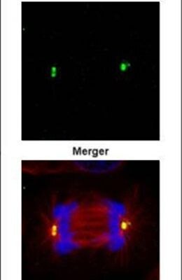
- Immunofluorescent analysis of gamma Tubulin in paraformaldehyde-fixed 293T cells using a gamma Tubulin polyclonal antibody (Product # PA5-34815) (Green) at a 1:500 dilution. Alpha-tubulin filaments were labeled with Product # PA5-29281 (Red) at a 1:500.
- Submitted by
- Invitrogen Antibodies (provider)
- Main image
- Experimental details
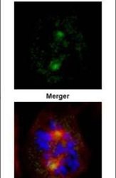
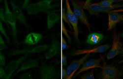
- gamma Tubulin Polyclonal Antibody detects gamma Tubulin protein at centrosome by immunofluorescent analysis. Sample: HeLa cells were fixed in ice-cold MeOH for 5 min. Green: gamma Tubulin stained by gamma Tubulin Polyclonal Antibody (Product # PA5-34815) diluted at 1:1,000. Red: alpha Tubulin, stained by alpha Tubulin Polyclonal Antibody [GT114] (Product # MA5-31466) diluted at 1:500. Blue: Fluoroshield with DAPI .

- Submitted by
- Invitrogen Antibodies (provider)
- Main image

- Experimental details
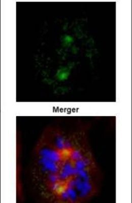
- gamma Tubulin Polyclonal Antibody detects gamma Tubulin protein at centrosome by immunofluorescent analysis. Sample: HeLa cells were fixed in ice-cold MeOH for 5 min. Green: gamma Tubulin stained by gamma Tubulin Polyclonal Antibody (Product # PA5-34815) diluted at 1:1,000. Red: alpha Tubulin, stained by alpha Tubulin Polyclonal Antibody [GT114] (Product # MA5-31466) diluted at 1:500. Blue: Fluoroshield with DAPI .

Supportive validation
- Submitted by
- Invitrogen Antibodies (provider)
- Main image

- Experimental details
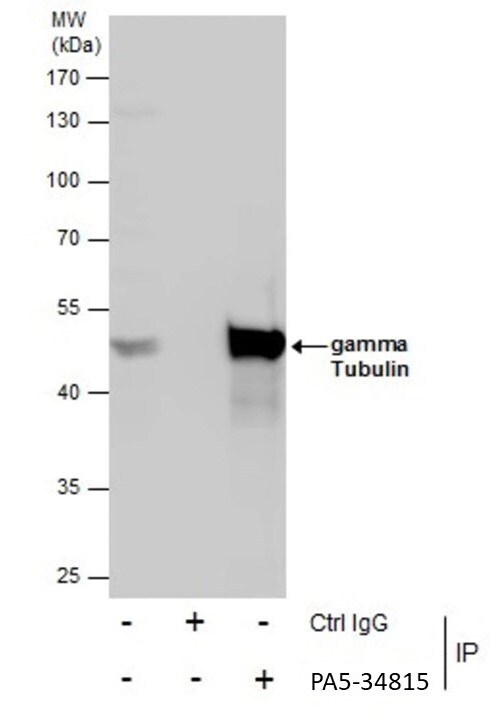
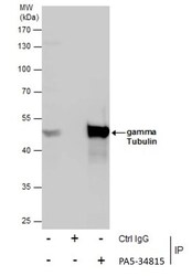
- Immunoprecipitation of gamma Tubulin was performed in Jurkat whole cell extracts using 5 µg of gamma Tubulin Polyclonal Antibody (Product # PA5-34815). Samples were transferred to a membrane and probed with gamma Tubulin Polyclonal Antibody as a primary antibody and an HRP-conjugated anti-Rabbit IgG was used as a secondary antibody.
Supportive validation
- Submitted by
- Invitrogen Antibodies (provider)
- Main image

- Experimental details
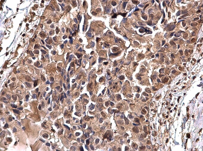
- gamma Tubulin Polyclonal Antibody detects gamma Tubulin protein at nucleus on human breast carcinoma by immunohistochemical analysis. Sample: Paraffin-embedded human breast carcinoma. Gamma Tubulin Polyclonal Antibody (Product # PA5-34815) dilution: 1:500. Antigen Retrieval: EDTA based buffer, pH 8.0, 15 min.
Supportive validation
- Submitted by
- Invitrogen Antibodies (provider)
- Main image
- Experimental details
- Immunoprecipitation of gamma Tubulin was performed in Jurkat whole cell extracts using 5 µg of gamma Tubulin Polyclonal Antibody (Product # PA5-34815). Samples were transferred to a membrane and probed with gamma Tubulin Polyclonal Antibody as a primary antibody and an HRP-conjugated anti-Rabbit IgG was used as a secondary antibody.

- Submitted by
- Invitrogen Antibodies (provider)
- Main image

- Experimental details
- Fig. 1 PPP1R2 affects centrosome number through interaction with AURKA and PP1. a-b Schematic of constructs used for transfection and representative transfection. c-i ARPE-19 cells were transfected either singly or in combination with plasmids expressing PPP1R2, AURKA, PP1 and empty vector as control (FLAG). Transfected cells were stained for gamma-tubulin (green) and alpha-tubulin (red) and centrosomes were counted in a minimum of 100 cells for each treatment group in three replicates. Insets magnify cellular regions containing centrosomes. Size bar equals 10 mum. j Graphical representation of the frequency of supernumerary centrosomes in cells transfected with each of the indicated plasmids individually or in combination. Statistically significant differences ( p < 0.05) between groups are indicated by differing letter notations above the bars and error bars represent standard error of the mean. Statistically significant differences are indicated with asterisks (* = p
