Explore
 Validate
 Learn
Learn Western blot
 Immunocytochemistry
Antibody data
- Antibody Data
- Antigen structure
- References [2]
- Comments [0]
- Validations
- Western blot [1]
- Immunocytochemistry [1]
- Immunohistochemistry [1]
Submit
Validation data
Reference
Comment
Report error
- Product number
- AMAb90544 - Provider product page

- Provider
- Atlas Antibodies
- Proper citation
- Atlas Antibodies Cat#AMAb90544, RRID:AB_2665581
- Product name
- Anti-SIX1
- Antibody type
- Monoclonal
- Description
- Monoclonal Antibody against Human SIX1, Clone ID: CL0185, Gene description: SIX homeobox 1, Alternative Gene Names: DFNA23, Validated applications: ICC, IHC, WB, Uniprot ID: Q15475, Storage: Store at +4°C for short term storage. Long time storage is recommended at -20°C.
- Reactivity
- Human
- Host
- Mouse
- Conjugate
- Unconjugated
- Isotype
- IgG
- Antibody clone number
- CL0185
- Vial size
- 100 µl
- Concentration
- 1.0 mg/ml
- Storage
- Store at +4°C for short term storage. Long time storage is recommended at -20°C.
- Handling
- The antibody solution should be gently mixed before use.
Submitted references Critical roles of FGF, RA, and WNT signalling in the development of the human otic placode and subsequent lineages in a dish.
Lateral line placodes of aquatic vertebrates are evolutionarily conserved in mammals.
Saeki T, Yoshimatsu S, Ishikawa M, Hon CC, Koya I, Shibata S, Hosoya M, Saegusa C, Ogawa K, Shin JW, Fujioka M, Okano H
Regenerative therapy 2022 Jun;20:165-186
Regenerative therapy 2022 Jun;20:165-186
Lateral line placodes of aquatic vertebrates are evolutionarily conserved in mammals.
Washausen S, Knabe W
Biology open 2018 Jun 19;7(6)
Biology open 2018 Jun 19;7(6)
No comments: Submit comment
Enhanced validation
- Submitted by
- Atlas Antibodies (provider)
- Enhanced method
- Genetic validation
- Main image
- Experimental details
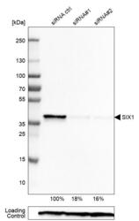
- Western blot analysis in Rh30 cells transfected with control siRNA, target specific siRNA probe #1 and #2, using Anti-SIX1 antibody. Remaining relative intensity is presented. Loading control: Anti-PPIB.
- Sample type
- Human
- Protocol
- Protocol

Supportive validation
- Submitted by
- Atlas Antibodies (provider)
- Main image

- Experimental details
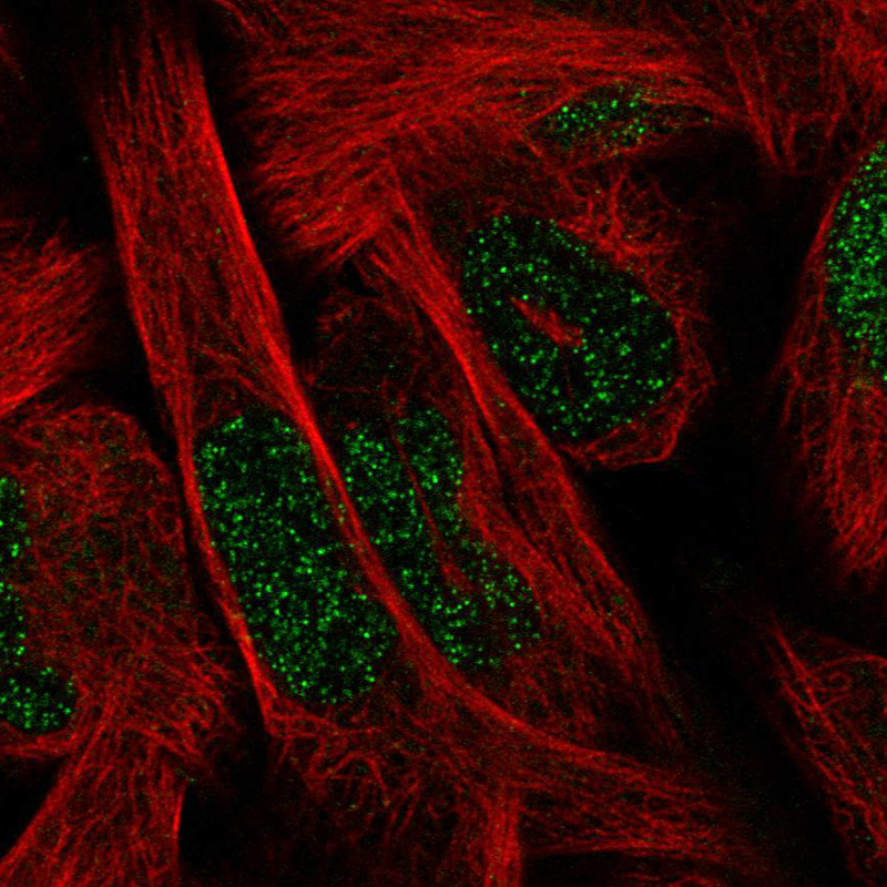
- Immunofluorescence staining in RH30 cell line with Anti-SIX1 monoclonal antibody, showing spotty nuclear staining in green. Microtubule probes are visualized in red (where available).
- Sample type
- Human
Supportive validation
- Submitted by
- Atlas Antibodies (provider)
- Enhanced method
- Orthogonal validation
- Main image
- Experimental details
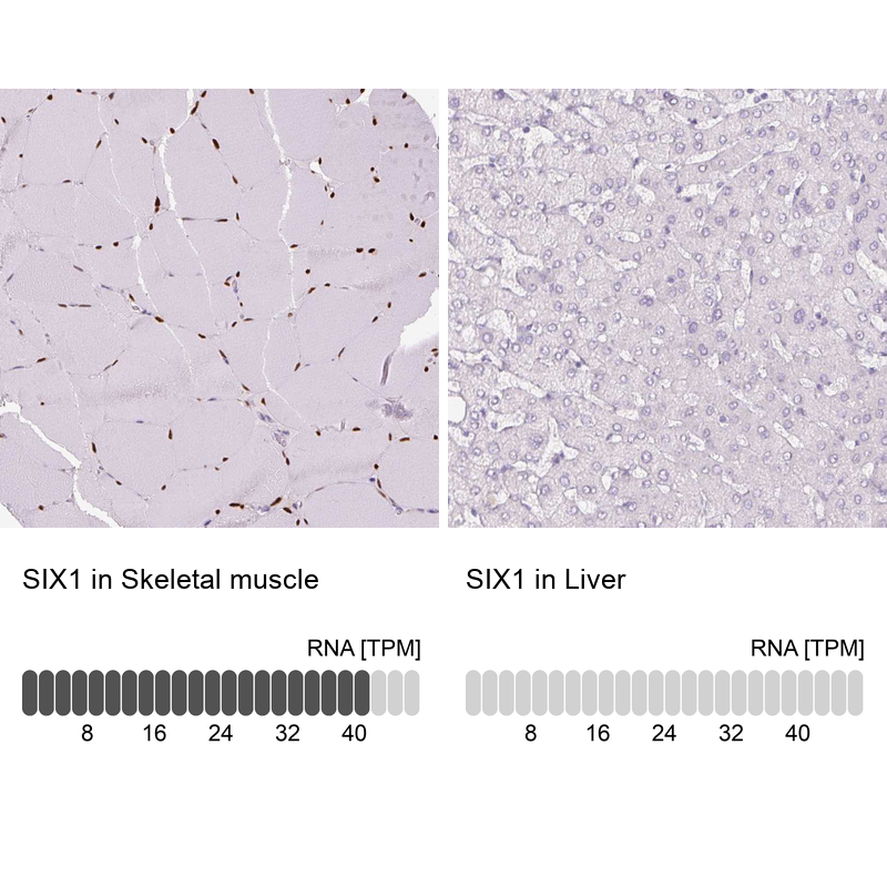
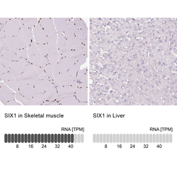
- Immunohistochemistry analysis in human skeletal muscle and liver tissues using AMAb90544 antibody. Corresponding SIX1 RNA-seq data are presented for the same tissues.
- Sample type
- Human
- Protocol
- Protocol