 Explore
 Validate
 Learn
Learn Western blot
Antibody data
- Antibody Data
- Antigen structure
- References [3]
- Comments [0]
- Validations
- Western blot [1]
- Immunohistochemistry [1]
Submit
Validation data
Reference
Comment
Report error
- Product number
- PAB3322 - Provider product page

- Provider
- Abnova Corporation
- Proper citation
- Abnova Corporation Cat#PAB3322, RRID:AB_1581922
- Product name
- WEE1 polyclonal antibody
- Antibody type
- Polyclonal
- Description
- Rabbit polyclonal antibody raised against synthetic peptide of WEE1.
- Storage
- Store at 4°C. For long term storage store at -20°C.Aliquot to avoid repeated freezing and thawing.
Submitted references Human wee1 kinase is directly transactivated by and increased in association with c-Fos/AP-1: rheumatoid synovial cells overexpressing these genes go into aberrant mitosis.
Depletion of Wee-1 kinase is necessary for both human immunodeficiency virus type 1 Vpr- and gamma irradiation-induced apoptosis.
Inhibition of proteasome-dependent degradation of Wee1 in G2-arrested Hep3B cells by TGF beta 1.
Kawasaki H, Komai K, Nakamura M, Yamamoto E, Ouyang Z, Nakashima T, Morisawa T, Hashiramoto A, Shiozawa K, Ishikawa H, Kurosaka M, Shiozawa S
Oncogene 2003 Oct 9;22(44):6839-44
Oncogene 2003 Oct 9;22(44):6839-44
Depletion of Wee-1 kinase is necessary for both human immunodeficiency virus type 1 Vpr- and gamma irradiation-induced apoptosis.
Yuan H, Xie YM, Chen IS
Journal of virology 2003 Feb;77(3):2063-70
Journal of virology 2003 Feb;77(3):2063-70
Inhibition of proteasome-dependent degradation of Wee1 in G2-arrested Hep3B cells by TGF beta 1.
Hashimoto O, Ueno T, Kimura R, Ohtsubo M, Nakamura T, Koga H, Torimura T, Uchida S, Yamashita K, Sata M
Molecular carcinogenesis 2003 Apr;36(4):171-82
Molecular carcinogenesis 2003 Apr;36(4):171-82
No comments: Submit comment
Supportive validation
- Submitted by
- Abnova Corporation (provider)
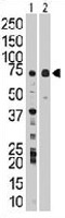
- Main image

- Experimental details
- The WEE1 polyclonal antibody (Cat # PAB3322) is used in Western blot to detect WEE1 in Jurkat cell lysate (Lane 1) and mouse liver tissue lysate (Lane 2) .
Supportive validation
- Submitted by
- Abnova Corporation (provider)
- Main image
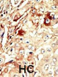
- Experimental details
- Formalin-fixed and paraffin-embedded human hepatocellular carcinoma tissue reacted with WEE1 polyclonal antibody (Cat # PAB3322) , which was peroxidase-conjugated to the secondary antibody, followed by AEC staining. This data demonstrates the use of this antibody for immunohistochemistry; clinical relevance has not been evaluated. HC = hepatocarcinoma.
- Validation comment
- Immunohistochemistry (Formalin/PFA-fixed paraffin-embedded sections)
