 Explore
 Validate
 Learn
Learn Western blot
 ELISA
ELISAAntibody data
- Antibody Data
- Antigen structure
- References [0]
- Comments [0]
- Validations
- Western blot [2]
- Immunocytochemistry [1]
- Flow cytometry [1]
- Other assay [12]
Submit
Validation data
Reference
Comment
Report error
- Product number
- 51-0700 - Provider product page

- Provider
- Invitrogen Antibodies
- Product name
- 14-3-3 Pan Polyclonal Antibody
- Antibody type
- Polyclonal
- Antigen
- Synthetic peptide
- Description
- This antibody is broadly reactive with members of the 14-3-3 family of proteins (~29-32 kDa). With the exception of a single conserved amino acid change, this sequence is identical in the delta and zeta isoforms. Three and four conserved amino acid differences are found in the tau and gamma isoforms, respectively. Based on the sequence homology to the immunizing peptide, this the antibody could recognize the protein from C. elegans.
- Reactivity
- Human, Mouse, Rat, Bovine, Xenopus
- Host
- Rabbit
- Isotype
- IgG
- Vial size
- 200 µg
- Concentration
- 0.25 mg/mL
- Storage
- Maintain refrigerated at 2-8°C for up to 1 month. For long term storage store at -20°C
No comments: Submit comment
Supportive validation
- Submitted by
- Invitrogen Antibodies (provider)
- Main image
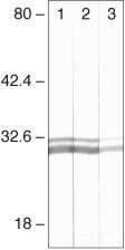
- Experimental details
- Western blot analysis of HeLa cells using Rb anti-14-3-3 (Product # 51-0700). Lane 1: 1 µg/mL, Lane 2: 0.5, g/mL, Lane 3: 0.1 µg/mL

- Submitted by
- Invitrogen Antibodies (provider)
- Main image
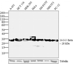
- Experimental details
- Western blot analysis was performed on whole cell extracts (30 µg) of A-431 (Lane 1), HCT 116 (Lane 2), HEK-293 (Lane 3), HeLa (Lane 4), SH-SY5Y (Lane 5), NIH/3T3 (Lane 6) and PC-12 (Lane 7). The blots were probed with Anti- 14-3-3 theta Rabbit Polyclonal Antibody (Product # 51-0700, 2 µg/mL) and detected by chemiluminescence using Goat anti-Rabbit IgG (H+L) Superclonal™ Secondary Antibody, HRP conj µgate (Product # A27036, 0.4 µg/mL, 1:2500 dilution). A ~ 28 kDa band corresponding to 14-3-3 theta was observed across cell lines tested. Known quantity of protein samples were electrophoresed using Novex® NuPAGE® 4-12 % Bis-Tris gel (Product # NP0321BOX), XCell SureLock™ Electrophoresis System (Product # EI0002) and Novex® Sharp Pre-Stained Protein Standard (Product # LC5800). Resolved proteins were then transferred onto a nitrocellulose membrane with iBlot® 2 Dry Blotting System (Product # IB21001). The membrane was probed with the relevant primary and secondary Antibody following blocking with 5 % skimmed milk. Chemiluminescent detection was performed using Pierce™ ECL Western Blotting Substrate (Product # 32106).

Supportive validation
- Submitted by
- Invitrogen Antibodies (provider)
- Main image

- Experimental details
- Immunofluorescence analysis of 14-3-3 theta was performed using 70% confluent log phase MCF-7 cells. The cells were fixed with 4% paraformaldehyde for 10 minutes, permeabilized with 0.1% Triton™ X-100 for 10 minutes, and blocked with 1% BSA for 1 hour at room temperature. The cells were labeled with 14-3-3 theta Rabbit Polyclonal Antibody (Product # 51-0700) at 2 µg/mL in 0.1% BSA and incubated for 3 hours at room temperature and then labeled with Goat anti-Rabbit IgG (H+L) Superclonal™ Secondary Antibody, Alexa Fluor® 488 conj µgate (Product # A27034) at a dilution of 1:2000 for 45 minutes at room temperature (Panel a: green). Nuclei (Panel b: blue) were stained with SlowFade® Gold Antifade Mountant with DAPI (Product # S36938). F-actin (Panel c: red) was stained with Alexa Fluor® 555 Rhodamine Phalloidin (Product # R415, 1:300). Panel d represents the merged image showing cytoplasmic localization. Panel e shows the no primary antibody control. The images were captured at 60X magnification.

Supportive validation
- Submitted by
- Invitrogen Antibodies (provider)
- Main image
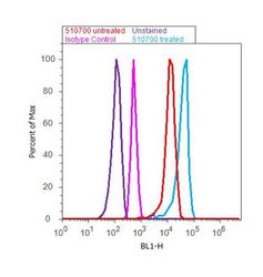
- Experimental details
- Flow Cytometry analysis of 14-3-3 theta was done on MCF7 cells (untreated, red histogram) and MCF7 cells treated with 100uM Etoposide for 6 hours (blue histogram). Cells were fixed with 70% ethanol for 10 minutes, permeabilized with 0.25% Triton™ X-100 for 20 minutes, and blocked with 5% BSA for 30 minutes at room temperature. Cells were labeled with 14-3-3 theta Rabbit Polyclonal Antibody (Product # 51-0700) or with rabbit isotype control (pink histogram) at 3-5 µg/million cells in 2.5% BSA. After incubation at room temperature for 2 hours, the cells were labeled with Alexa Fluor® 488 Goat Anti-Rabbit Secondary Antibody (Product # A11008) at a dilution of 1:400 for 30 minutes at room temperature. The representative 10, 000 cells were acquired and analyzed for each sample using an Attune® Acoustic Focusing Cytometer. The purple histogram represents unstained control cells.

Supportive validation
- Submitted by
- Invitrogen Antibodies (provider)
- Main image

- Experimental details
- NULL

- Submitted by
- Invitrogen Antibodies (provider)
- Main image

- Experimental details
- NULL

- Submitted by
- Invitrogen Antibodies (provider)
- Main image

- Experimental details
- NULL

- Submitted by
- Invitrogen Antibodies (provider)
- Main image

- Experimental details
- NULL

- Submitted by
- Invitrogen Antibodies (provider)
- Main image

- Experimental details
- NULL

- Submitted by
- Invitrogen Antibodies (provider)
- Main image

- Experimental details
- NULL

- Submitted by
- Invitrogen Antibodies (provider)
- Main image

- Experimental details
- Figure 3 Effects of Tctp mutation on Rheb and 14-3-3s. ( a - d ) Clones of Tctp h59 mutant cells in eye disc. ( e - h ) Clones of Tctp h59 mutant cells in wing disc. These clones are marked by the absence of Arm-lacZ. In both organs, Tctp mutant clones show reduction of Rheb ( c , g ) but not 14-3-3s ( d , h ). Red rectangles in a and e indicate the enlarged regions shown in b - d and f - h respectively. Scale bar, 100 mum ( a - h ).

- Submitted by
- Invitrogen Antibodies (provider)
- Main image

- Experimental details
- Figure 5 14-3-3s are required for the interaction between Tctp and Rheb and organ development. ( a ) Knockdown of 14-3-3s in S2 cells shows depletion of 14-3-3s levels after 6 days of RNAi treatment. ei, zetai and ei+zetai indicate RNAi for 14-3-3e, 14-3-3zeta and both isoforms, respectively. Levels of Tctp and Rheb were not noticeably affected by 14-3-3 depletion. The treatment performed to detect Tctp, Rheb, and actin level was double knockdown of both isoforms of 14-3-3. ( b ) 14-3-3 depletion affects the interaction between V5-Tctp and V5myc-Rheb after 6 days of RNAi treatment. Anti-Myc antibody was used for IP. Tctp was detected by anti-Tctp antibody. Depletion of both isoforms abolished the Tctp-Rheb interaction. ( c - j ) Double knockdown of 14-3-3s causes loss of targeted tissues. ey-Gal4 control ( c ) 14-3-3e RNAi ( d ) and 14-3-3zeta RNAi ( e ) show normal pattern of eye-head in pupae. Knockdown of both 14-3-3 isoforms causes pupal lethality with loss of eye and head ( f ). ( g - j ) Pattern of Elav staining in larval eye discs from indicated genotypes. Knockdown of 14-3-3e or 14-3-3zeta has no obvious effect on eye disc development ( h , i ). Knockdown of both isoforms results in small disc (the area marked with dotted line) with no retinal differentiation ( j ). ( k - n ) Double knockdown of 14-3-3s results in reduced proliferation. MS1096-Gal4 control ( k ) 14-3-3e RNAi ( l ) and 14-3-3zeta RNAi ( m ) show normal level of PH3 staining in wing disc. Knockdown of b

- Submitted by
- Invitrogen Antibodies (provider)
- Main image

- Experimental details
- Figure 6 Effects of 14-3-3 RNAi on Tor targets and the suppression by CycE. ( a ) Effects of 14-3-3 RNAi on S6k and Thor. Either one of 14-3-3 isoforms or both 14-3-3s were knocked down in S2 cells. Single or double knockdown of 14-3-3s reduced the level of pS6k (Thr 398) but does not affect the level of S6k protein. Knockdown of both 14-3-3s, but not one isoform, causes slight reduction of Thor. In contrast, pThor (Thr 37/46) is strongly reduced by single or double knockdown of 14-3-3s. ( b - d ) Loss of 14-3-3s results in reduction of CycE level. ( b ) 14-3-3e mutant clones induced by ey-flp are marked by the absence of GFP. Mutant clones show strong reduction of CycE ( c ). ( e - g ) 14-3-3zeta mutant clones induced by hs-flp are marked by the absence of Arm-lacZ. CycE is reduced in mutant clones ( f ). ( h - m ) Effects of CycE in eye. Control with one copy of ey-Gal4 ( h ) or overexpression of CycE ( i ) shows normal eye. ( j ) Double knockdown of Tctp and 14-3-3 results in small and rough eye phenotype (Tctp RNAi/ CycE GFP ; 14-3-3 RNAi/+ were scored. ( o - r ) CycE suppresses the wing pheno

- Submitted by
- Invitrogen Antibodies (provider)
- Main image

- Experimental details
- Figure 9 ( A )Western blotting of differentially expressed proteins in PP2C9TL and WT at 10 DAF stage. The top photo shows the SDS-PAGE separation of two protein samples used as loading control. The bottom photo shows gel transferred onto nitrocellulose membrane for western blotting to detect actin, anti-Flag- PP2C9 , and 14-3-3 protein for WT and PP2C9TL at the 10 DAF stage. ( B ) Co-immunoprecipitation (Co-IP) assays of PP2C9 associated protein analyzed by SDS-PAGE. Protein molecular weight marker, negative control without antibodies, WT protein, and the third is the PP2C9TL protein. 2.8. Identification of PP2C9 Associated Protein by Co-Immunoprecipitation (CO-IP) and Mass Spectrometry.

- Submitted by
- Invitrogen Antibodies (provider)
- Main image

- Experimental details
- Figure 3 Confirmation of interactions between Parkin and selected target proteins in human and murine platelets. ( A ) Immunoprecipitation of each specific antibody in pooled human DM platelets (4 HC, 5DM1, and 6DM2). We incubated 500 mug protein lysates incubated with specific antibodies overnight at 4 degC with 10% input as control. G1 represents interacting protein groups from LC-MS/MS results. G2 represents the non-interacting proteins group in LC-MS/MS results. G3 represents protein that interacted with Parkin and that were not found in the LC-MS/MS results. ( B ) Immunoprecipitation of each specific antibody in pooled murine platelets (3 WT and 5DM). We incubated 500 mug protein lysates incubated with specific antibodies overnight at 4 degC with 10% input as control.

- Submitted by
- Invitrogen Antibodies (provider)
- Main image

- Experimental details
- Figure 4. Drosophila 14-3-3 proteins interact with Dlg in the wing disc epithelium. (A) Coimmunoprecipitation of endogenous 14-3-3 proteins and Dlg from wild-type wing disc samples. Western blots were probed with anti-14-3-3 antibody (upper lanes) and anti-Dlg antibody (lower lanes) in input and immunoprecipitates pulled down by anti-Dlg antibody. IP, immunoprecipitation. (B-G) PLA indicates close proximity or interactions of two proteins. PLA between Scrib and Dlg in control ( nub-Gal4/ +; B) and dlg -knockdown ( nub-Gal4>dlg-RNAi ; C) wing discs. (D) Quantification of the number of PLA (Scrib/Dlg) spots for control ( n = 6) and dlg-RNAi ( n = 5) wing discs. PLA between 14-3-3 and Dlg in control (E) and dlg -knockdown (F) wing discs. (G) Quantification of the number of PLA (14-3-3/Dlg) spots for control ( n = 5) and dlg-RNAi ( n = 5) wing discs. Error bars are SD. **, P < 0.01 by Kolmogorov-Smirnov test. Note that PLA signals in MZ of the disc proper diminished in dlg -RNAi wing discs, but not in PE where nub-Gal4 is not expressed. PE, peripodial epithelium; MZ, mitotic zone. (H and I) Subcellular localization of 14-3-3 proteins detected by anti-14-3-3 antibody staining. 14-3-3 proteins localize to the apical cortex and nucleus during interphase (H) and cytosol, including spindle microtubules (alpha-tubulin), during mitosis (I). Yellow arrows indicate metaphase cells. Scale bars: 5 um.
