Explore
 Validate
 Learn
Learn Western blot
Antibody data
- Antibody Data
- Antigen structure
- References [1]
- Comments [0]
- Validations
- Western blot [5]
- Immunocytochemistry [5]
- Immunoprecipitation [2]
- Immunohistochemistry [3]
- Other assay [3]
Submit
Validation data
Reference
Comment
Report error
- Product number
- PA5-27317 - Provider product page

- Provider
- Invitrogen Antibodies
- Product name
- 14-3-3 zeta Polyclonal Antibody
- Antibody type
- Polyclonal
- Antigen
- Recombinant full-length protein
- Description
- Recommended positive controls: 293T, A431, H1299, HeLaS3, HepG2, Molt-4, Raji, NIH-3T3. Predicted reactivity: Mouse (99%), Rat (99%), Xenopus laevis (89%), Pig (99%), Chicken (99%), Sheep (99%), Rhesus Monkey (100%), Bovine (100%). Store product as a concentrated solution. Centrifuge briefly prior to opening the vial.
- Reactivity
- Human, Mouse, Rat
- Host
- Rabbit
- Isotype
- IgG
- Vial size
- 100 μL
- Concentration
- 0.4 mg/mL
- Storage
- Store at 4°C short term. For long term storage, store at -20°C, avoiding freeze/thaw cycles.
Submitted references 14-3-3ζ negatively regulates mitochondrial biogenesis in GBM residual cells.
Rajendra J, Ghorai A, Dutt S
Heliyon 2021 Nov;7(11):e08371
Heliyon 2021 Nov;7(11):e08371
No comments: Submit comment
Supportive validation
- Submitted by
- Invitrogen Antibodies (provider)
- Main image
- Experimental details
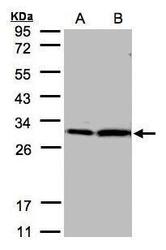
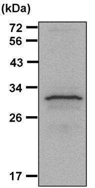
- Western Blot using 14-3-3 zeta Polyclonal Antibody (Product # PA5-27317). Sample (30 µg of whole cell lysate). A: Hep G2. B: MOLT4SDS PAGE. Z. 12% SDS PAGE.14-3-3 zeta Polyclonal Antibody (Product # PA5-27317) diluted at 1:2,000. The HRP-conjugated anti-rabbit IgG antibody was used to detect the primary antibody.
- Submitted by
- Invitrogen Antibodies (provider)
- Main image

- Experimental details
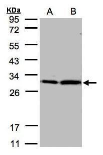
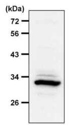
- Western Blot using 14-3-3 zeta Polyclonal Antibody (Product # PA5-27317). Sample (30 µg of whole cell lysate). Lane A: NIH-3T3. 12% SDS PAGE.14-3-3 zeta Polyclonal Antibody (Product # PA5-27317) diluted at 1:1,000. The HRP-conjugated anti-rabbit IgG antibody was used to detect the primary antibody.
- Submitted by
- Invitrogen Antibodies (provider)
- Main image
- Experimental details
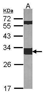
- Western blot analysis of 14-3-3 zeta was performed by loading 30 µg of THP-1 whole cell lysate per well onto an SDS-PAGE gel. Proteins were transferred to a PVDF membrane and blocked with 5% non-fat dry milk in TBST for 1 hour at room temperature. The membrane was probed with a 14-3-3 zeta polyclonal antibody (Product # PA5-27317) at a dilution of 1:1000 overnight at 4°C, washed in TBST, and probed with an HRP-conjugated goat anti-rabbit IgG secondary antibody at a dilution of 1:40,000 for 1 hour at room temperature. Detection was performed using ECL substrate. Data courtesy of the Innovators Program.

- Submitted by
- Invitrogen Antibodies (provider)
- Main image
- Experimental details
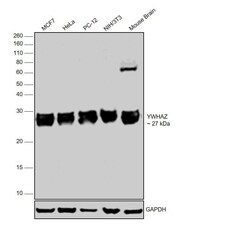
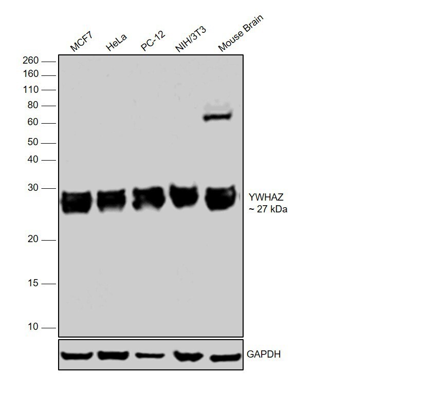
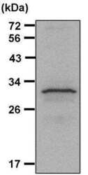
- Western blot was performed using Anti-14-3-3 zeta Polyclonal Antibody (Product # PA5-27317) and a 27 kDa band corresponding to YWHAZ was observed across all the cell lines and tissues tested. Whole cell extracts (25 µg lysate) of MCF7 (Lane 1), HeLa (Lane 2), PC-12 (Lane 3), NIH/3T3 (Lane 4) and tissue extracts of Mouse Brain (Lane 5) were electrophoresed using Novex® NuPAGE® 12 % Bis-Tris gel (Product # NP0342BOX). Resolved proteins were then transferred onto a nitrocellulose membrane (Product # IB23001) by iBlot® 2 Dry Blotting System (Product # IB21001). The blot was probed with the primary antibody (1:2000 dilution) and detected by chemiluminescence with Goat anti-Rabbit IgG (Heavy Chain), Superclonal™ Recombinant Secondary Antibody, HRP (Product # A27036, 1:4000 dilution) using the iBright FL 1000 (Product # A32752). Chemiluminescent detection was performed using Novex® ECL Chemiluminescent Substrate Reagent Kit (Product # WP20005).
- Submitted by
- Invitrogen Antibodies (provider)
- Main image

- Experimental details
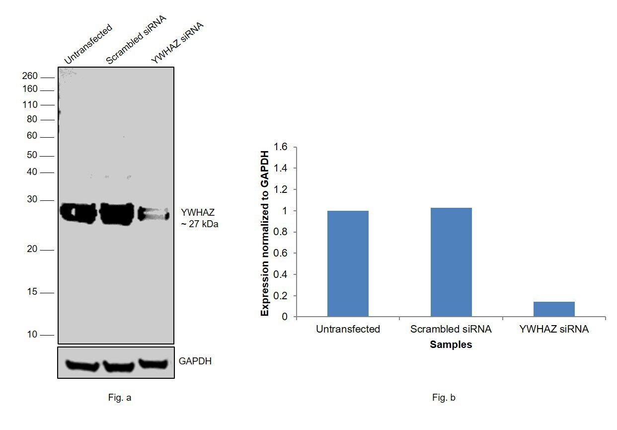
- Knockdown of 14-3-3 zeta was achieved by transfecting MCF7 with YWHAZ specific siRNAs (Silencer® select Product # s14972). Western blot analysis (Fig. a) was performed using whole cell extracts from the YWHAZ knockdown cells (Lane 3), non-specific scrambled siRNA transfected cells (Lane 2) and untransfected cells (Lane 1). The blot was probed with 14-3-3 zeta Polyclonal Antibody (Product # PA5-27317, 1:2,000 dilution) and Goat anti-Rabbit IgG (Heavy Chain), Superclonal™ Recombinant Secondary Antibody, HRP (Product # A27036, 0.25 µg/mL, 1:4,000 dilution). Densitometric analysis of this western blot is shown in histogram (Fig. b). Decrease in signal upon siRNA mediated knock down confirms that antibody is specific to YWHAZ.
Supportive validation
- Submitted by
- Invitrogen Antibodies (provider)
- Main image
- Experimental details
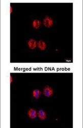
- Immunofluorescent analysis of 14-3-3 zeta in paraformaldehyde-fixed HeLaS3 cells using a 14-3-3 zeta polyclonal antibody (Product # PA5-27317) at a 1:100 dilution.

- Submitted by
- Invitrogen Antibodies (provider)
- Main image

- Experimental details
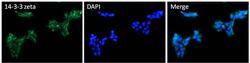
- Immunofluorescent analysis of 14-3-3 zeta (green) HEK293T cells. Cells fixed with 4% formaldehyde fixed were permeabilized and blocked with 1X PBS containing 5% BSA and 0.3% Triton X-100 for 1 hour at room temperature. Cells were probed with a 14-3-3 zeta ppolyclonal antibody (Product # PA5-27317) at a dilution of 1:100 overnight at 4°C in 1X PBS containing 1% BSA and 0.3% Triton X-100, washed with 1X PBS, and incubated with fluorophore-conjugated goat anti-rabbit IgG secondary antibody at a dilution of 1:200 for 1 hour at room temperature. Nuclei (blue) were stained with DAPI. Images were taken on a Leica DM1000 microscope at 40X magnification. Data courtesy of the Innovators Program.

- Submitted by
- Invitrogen Antibodies (provider)
- Main image

- Experimental details
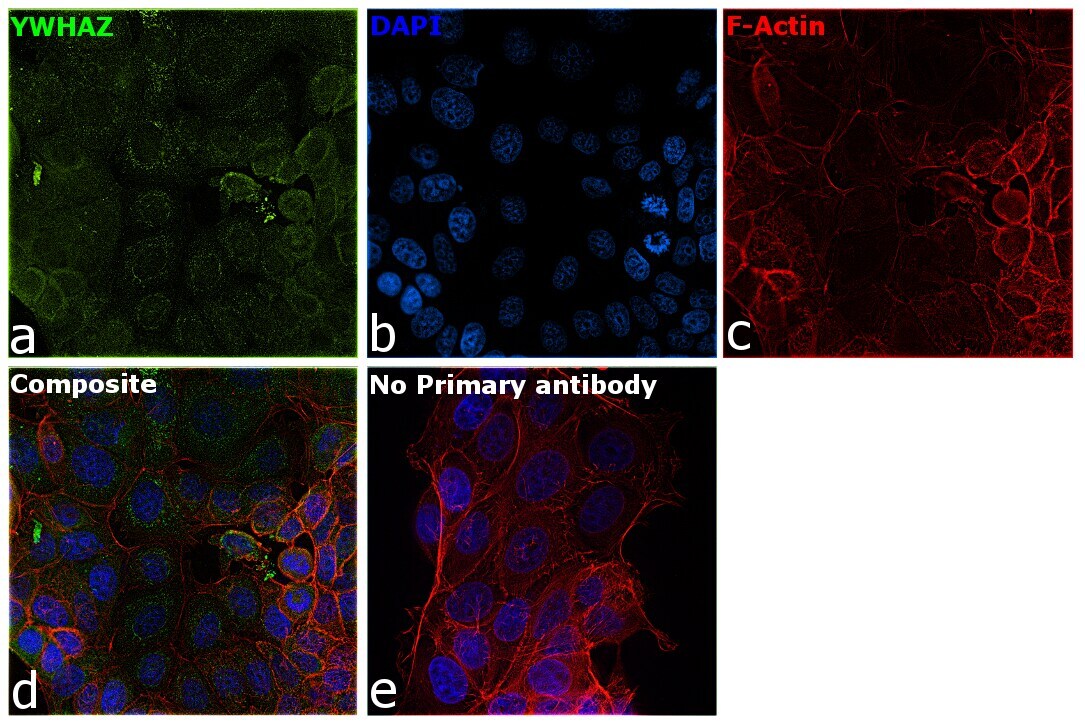
- Immunofluorescence analysis of YWHAZ was performed using 70% confluent log phase MCF7 cells. The cells were fixed with 4% Paraformaldehyde for 10 minutes, permeabilized with 0.1% Triton™ X-100 for 10 minutes, and blocked with 2% BSA for 10 minutes at room temperature. The cells were labeled with 14-3-3 zeta Polyclonal Antibody (Product # PA5-27317) at 1:200 dilution in 0.1% BSA, incubated at 4 degree celsius overnight and then labeled with Goat anti-Rabbit IgG (H+L) Highly Cross-Adsorbed Secondary Antibody, Alexa Fluor Plus 488 (Product # A32731, 1:2000 dilution) for 45 minutes at room temperature (Panel a: Green). Nuclei (Panel b: Blue) were stained with SlowFade® Gold Antifade Mountant with DAPI (Product # S36938). F-actin (Panel c: Red) was stained with Rhodamine Phalloidin (Product # R415, 1:300). Panel d represents the merged image showing Cytoplasmic and endoplasmic reticulum like localization of YWHAZ. Panel e represents control cells with no primary antibody to assess background. The images were captured at 60X magnification.

- Submitted by
- Invitrogen Antibodies (provider)
- Main image

- Experimental details
- Immunofluorescence analysis of YWHAZ was performed using 70% confluent log phase MCF7 cells. The cells were fixed with 4% Paraformaldehyde for 10 minutes, permeabilized with 0.1% Triton™ X-100 for 10 minutes, and blocked with 2% BSA for 10 minutes at room temperature. The cells were labeled with 14-3-3 zeta Polyclonal Antibody (Product # PA5-27317) at 1:200 dilution in 0.1% BSA, incubated at 4 degree celsius overnight and then labeled with Goat anti-Rabbit IgG (H+L) Highly Cross-Adsorbed Secondary Antibody, Alexa Fluor Plus 488 (Product # A32731, 1:2000 dilution) for 45 minutes at room temperature (Panel a: Green). Nuclei (Panel b: Blue) were stained with SlowFade® Gold Antifade Mountant with DAPI (Product # S36938). F-actin (Panel c: Red) was stained with Rhodamine Phalloidin (Product # R415, 1:300). Panel d represents the merged image showing Cytoplasmic and endoplasmic reticulum like localization of YWHAZ. Panel e represents control cells with no primary antibody to assess background. The images were captured at 60X magnification.
- Submitted by
- Invitrogen Antibodies (provider)
- Main image
- Experimental details
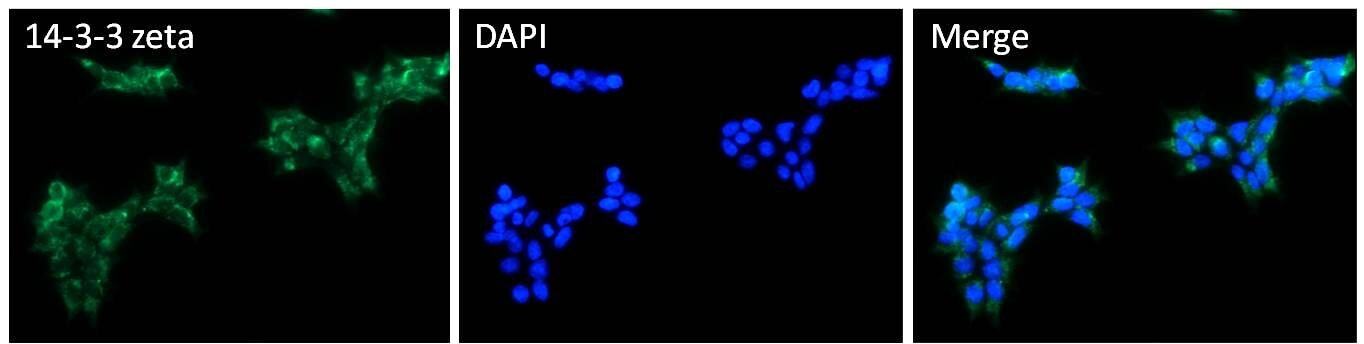
- Immunofluorescent analysis of 14-3-3 zeta (green) HEK293T cells. Cells fixed with 4% formaldehyde fixed were permeabilized and blocked with 1X PBS containing 5% BSA and 0.3% Triton X-100 for 1 hour at room temperature. Cells were probed with a 14-3-3 zeta ppolyclonal antibody (Product # PA5-27317) at a dilution of 1:100 overnight at 4°C in 1X PBS containing 1% BSA and 0.3% Triton X-100, washed with 1X PBS, and incubated with fluorophore-conjugated goat anti-rabbit IgG secondary antibody at a dilution of 1:200 for 1 hour at room temperature. Nuclei (blue) were stained with DAPI. Images were taken on a Leica DM1000 microscope at 40X magnification. Data courtesy of the Innovators Program.
Supportive validation
- Submitted by
- Invitrogen Antibodies (provider)
- Main image

- Experimental details
- Immunoprecipitation of 14-3-3 zeta was performed on THP-1 cells. Antigen-antibody complexes were formed by incubating 400ug of THP-1 whole cell lysate (in 400ul volume) with 5ul of a 14-3-3 zeta polyclonal antibody (Product # PA5-27317) overnight at 4¼C. The immune complexes were captured on 30ul of protein G agarose, washed extensively, and eluted with 6X non-reducing Laemmli buffer. Samples were resolved on a 10% SDS-PAGE, transferred to a PVDF membrane, and blocked with 5% milk in TBST for 1 hour at room temperature. The membrane was probed with a 14-3-3 zeta polyclonal antibody (Product # PA5-27317) at a dilution of 1:2000 overnight at 4¡C, washed in TBST, and probed with an HRP-conjugated goat anti-rabbit IgG secondary antibody at a dilution of 1:40,000 for 1 hour at room temperature. Chemiluminescent detection was performed using ECL substrate. Data courtesy of the Innovators Program.

- Submitted by
- Invitrogen Antibodies (provider)
- Main image

- Experimental details
- Immunoprecipitation of 14-3-3 zeta was performed on THP-1 cells. Antigen-antibody complexes were formed by incubating 400 µg of THP-1 whole cell lysate (in 400 µL volume) with 5 µL of a 14-3-3 zeta polyclonal antibody (Product # PA5-27317) overnight at 4°C. The immune complexes were captured on 30 µL of protein G agarose, washed extensively, and eluted with 6X non-reducing Laemmli buffer. Samples were resolved on a 10% SDS-PAGE, transferred to a PVDF membrane, and blocked with 5% milk in TBST for 1 hour at room temperature. The membrane was probed with a 14-3-3 zeta polyclonal antibody (Product # PA5-27317) at a dilution of 1:2000 overnight at 4°C, washed in TBST, and probed with an HRP-conjugated goat anti-rabbit IgG secondary antibody at a dilution of 1:40,000 for 1 hour at room temperature. Chemiluminescent detection was performed using ECL substrate. Data courtesy of the Innovators Program.

Supportive validation
- Submitted by
- Invitrogen Antibodies (provider)
- Main image
- Experimental details
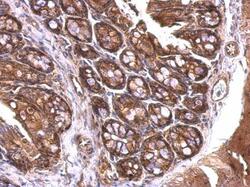
- 14-3-3 zeta Polyclonal Antibody detects 14-3-3 zeta protein at cytosol on mouse colon by immunohistochemical analysis. Sample: Paraffin-embedded mouse colon.14-3-3 zeta Polyclonal Antibody (Product # PA5-27317) dilution: 1:500. Antigen Retrieval: EDTA based buffer, pH 8.0, 15 min.

- Submitted by
- Invitrogen Antibodies (provider)
- Main image
- Experimental details
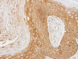
- Immunohistochemical analysis of paraffin-embedded Cal27 xenograft, using 14-3-3 zeta (Product # PA5-27317) antibody at 1:100 dilution. Antigen Retrieval: EDTA based buffer, pH 8.0, 15 min.

- Submitted by
- Invitrogen Antibodies (provider)
- Main image

- Experimental details
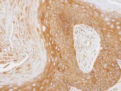
- Immunohistochemical analysis of paraffin-embedded Cal27 xenograft, using 14-3-3 zeta (Product # PA5-27317) antibody at 1:100 dilution. Antigen Retrieval: EDTA based buffer, pH 8.0, 15 min.
Supportive validation
- Submitted by
- Invitrogen Antibodies (provider)
- Main image
- Experimental details
- Immunoprecipitation of 14-3-3 zeta was performed on THP-1 cells. Antigen-antibody complexes were formed by incubating 400 µg of THP-1 whole cell lysate (in 400 µL volume) with 5 µL of a 14-3-3 zeta polyclonal antibody (Product # PA5-27317) overnight at 4°C. The immune complexes were captured on 30 µL of protein G agarose, washed extensively, and eluted with 6X non-reducing Laemmli buffer. Samples were resolved on a 10% SDS-PAGE, transferred to a PVDF membrane, and blocked with 5% milk in TBST for 1 hour at room temperature. The membrane was probed with a 14-3-3 zeta polyclonal antibody (Product # PA5-27317) at a dilution of 1:2000 overnight at 4øC, washed in TBST, and probed with an HRP-conjugated goat anti-rabbit IgG secondary antibody at a dilution of 1:40,000 for 1 hour at room temperature. Chemiluminescent detection was performed using ECL substrate. Data courtesy of the Innovators Program.
- Submitted by
- Invitrogen Antibodies (provider)
- Main image
- Experimental details
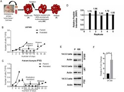
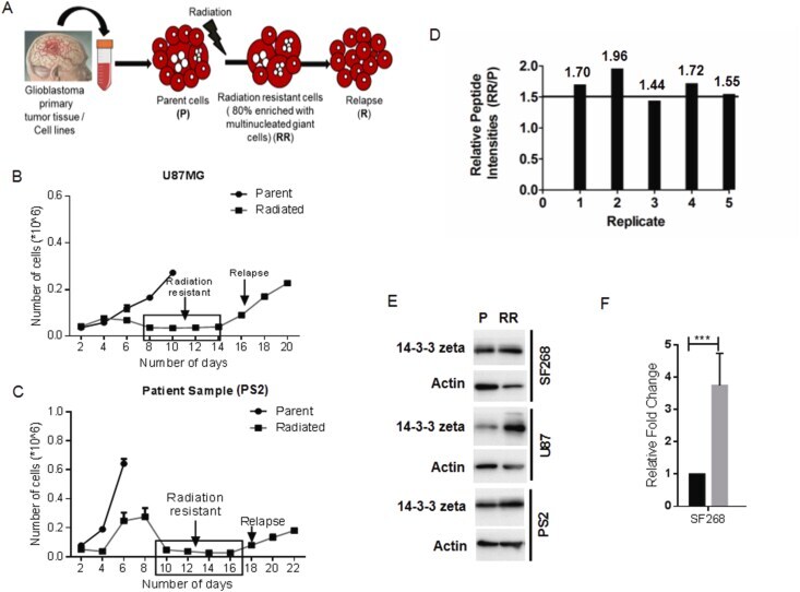
- Figure 1 Up-regulation of 14-3-3zeta in residual resistant disease cells. (A) Schematic diagram shows the in vitro radiation resistant model of Glioblastoma that captures residual disease cells (RR) as well as recurrent population (R) from the parent (P) population. (B) & (C) Cell proliferation assay using trypan blue staining showing the non-proliferative residual disease cells (radiation-resistant) and relapse in U87MG and patient sample 2 (PS2). (D) Graph shows relative peptide intensities of 14-3-3zeta in SF268 RR cells in five replicates of independent iTRAQ experiments. (E) Representative western blots showing the up-regulation of 14-3-3zeta in the RR cells of SF268, U87MG, and PS2. Beta-actin was used as a loading control (The uncropped original blots are provided as Supplementary Figure 1). (F) Real-time qPCR data showing the fold change of 14-3-3zeta gene expression in SF268 RR cells. Data represent mean +- SD of three independent experiments. *** denotes P
- Submitted by
- Invitrogen Antibodies (provider)
- Main image
- Experimental details
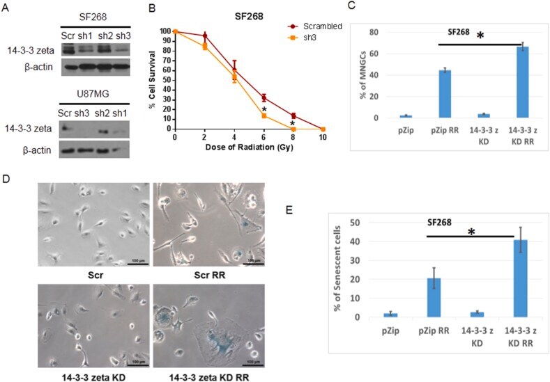
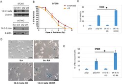
- Figure 2 14-3-3zeta knockdown increases MNGCs formation and radiation-induced senescence in RR cells. (A) Representative western blots showed the stable knockdown of 14-3-3zeta in SF268 and U87MG cells (The uncropped original blots are provided as Supplementary Figure 1). (B) Clonogenic radiation survival of SF268 cells after stable knockdown of 14-3-3zeta (C) Bar diagram depicts the percentage of MNGCs formed in RR population of SF268 after 14-3-3zeta knockdown. (D) Typical images of senescent cells showing beta-gal positivity in SF268 RR cells after 14-3-3zeta knockdown. Scale bar 50 mum. (E) Senescent positive cells were scored and plotted as the percentage of senescent positive cells. Data represent the mean +- SD of three independent experiments. *denotes P