Explore
 Validate
 Learn
Learn Western blot
Antibody data
- Antibody Data
- Antigen structure
- References [0]
- Comments [0]
- Validations
- Western blot [1]
- Immunohistochemistry [4]
Submit
Validation data
Reference
Comment
Report error
- Product number
- MA5-26828 - Provider product page

- Provider
- Invitrogen Antibodies
- Product name
- ALOX5 Monoclonal Antibody (OTI2C5)
- Antibody type
- Monoclonal
- Antigen
- Recombinant protein fragment
- Reactivity
- Human, Mouse, Rat
- Host
- Mouse
- Isotype
- IgG
- Antibody clone number
- OTI2C5
- Vial size
- 100 µL
- Concentration
- 1 mg/mL
- Storage
- -20° C, Avoid Freeze/Thaw Cycles
No comments: Submit comment
Supportive validation
- Submitted by
- Invitrogen Antibodies (provider)
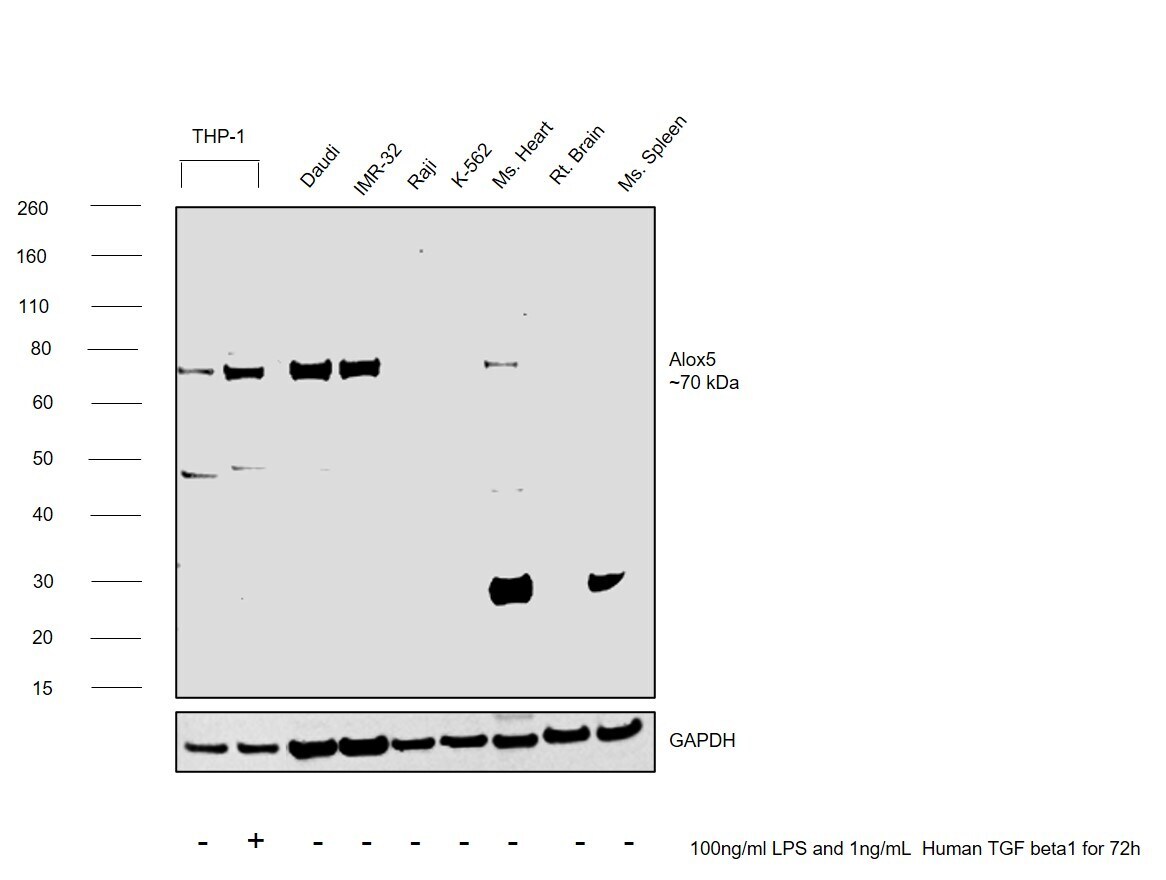
- Main image
- Experimental details
- Western blot was performed using Anti-ALOX5 Monoclonal Antibody (OTI2C5) (Product # MA5-26828) and a ~70 kDa band corresponding to Arachidonate 5-lipoxygenase was observed across cell lines and tissues tested. Whole cell extracts (30 µg lysate) of THP-1 (Lane 1), THP-1 treated with 100 ng/mL LPS and 1 ng/mL Hu. TGF beta 1 for 72h (Lane 2), Daudi (Lane 3), IMR-32 (Lane 4), Raji (Lane 5), K-562 (Lane 6), Mouse Heart (Lane 7), Rat Brain (Lane 8), Mouse Spleen (Lane 9) were electrophoresed using NuPAGE™ 4-12% Bis-Tris Protein Gel (Product # NP0321BOX). Resolved proteins were then transferred onto a nitrocellulose membrane (Product # IB23001) by iBlot® 2 Dry Blotting System (Product # IB21001). The blot was probed with the primary antibody (1:2000) and detected by chemiluminescence with Goat anti-Mouse IgG (H+L) Superclonal™ Recombinant Secondary Antibody, HRP (Product # A28177, 1:20000) using the iBright FL 1000 (Product # A32752). Chemiluminescent detection was performed using Novex® ECL Chemiluminescent Substrate Reagent Kit (Product # WP20005).Upregulation was observed in THP-1 cells treated with LPS and Human TGF beta 1. Increased pick up was observed in positive models (Daudi and IMR-32) compared to Raji and K-562.
Supportive validation
- Submitted by
- Invitrogen Antibodies (provider)
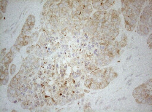
- Main image

- Experimental details
- Immunohistochemistry was performed on paraffin-embedded adenocarcinoma of human endometrium tissue. To expose target proteins, heat-induced epitope retrieval by 1mM EDTA in 10mM Tris buffer (pH8.5) at 120°C for 3 min. Following antigen retrieval, tissues were probed with a ALOX5 monoclonal antibody (Product # MA5-26828) at a dilution of 1:150.
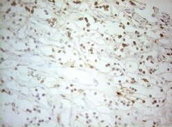
- Submitted by
- Invitrogen Antibodies (provider)
- Main image
- Experimental details
- Immunohistochemistry was performed on paraffin-embedded human tonsil tissue. To expose target proteins, heat-induced epitope retrieval by 1mM EDTA in 10mM Tris buffer (pH8.5) at 120°C for 3 min. Following antigen retrieval, tissues were probed with a ALOX5 monoclonal antibody (Product # MA5-26828) at a dilution of 1:150.

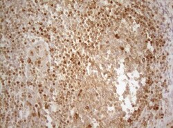
- Submitted by
- Invitrogen Antibodies (provider)
- Main image
- Experimental details
- Immunohistochemistry was performed on paraffin-embedded human lymphoma tissue. To expose target proteins, heat-induced epitope retrieval by 1mM EDTA in 10mM Tris buffer (pH8.5) at 120°C for 3 min. Following antigen retrieval, tissues were probed with a ALOX5 monoclonal antibody (Product # MA5-26828) at a dilution of 1:150.

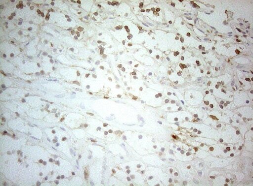
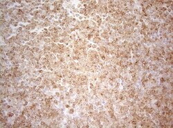
- Submitted by
- Invitrogen Antibodies (provider)
- Main image
- Experimental details
- Immunohistochemistry was performed on paraffin-embedded carcinoma of human kidney tissue. To expose target proteins, heat-induced epitope retrieval by 1mM EDTA in 10mM Tris buffer (pH8.5) at 120°C for 3 min. Following antigen retrieval, tissues were probed with a ALOX5 monoclonal antibody (Product # MA5-26828) at a dilution of 1:150.