 Explore
 Validate
 Learn
Learn Western blot
 ELISA
ELISAAntibody data
- Antibody Data
- Antigen structure
- References [1]
- Comments [0]
- Validations
- Western blot [2]
- Immunocytochemistry [3]
- Immunohistochemistry [2]
Submit
Validation data
Reference
Comment
Report error
- Product number
- MA5-38050 - Provider product page

- Provider
- Invitrogen Antibodies
- Product name
- ALOX5 Recombinant Rabbit Monoclonal Antibody (5C5Z2)
- Antibody type
- Monoclonal
- Antigen
- Synthetic peptide
- Description
- Positive Samples: Raji, Rat lung Immunogen sequence: CYRWITGDVE VVLRDGRAKL ARDDQIHILK QHRRKELETR QKQYRWMEWN PGFPLSIDAK CHKDLPRDIQ FDSEKGVDFV LNYSKAMENL FINRFMHMFQ S
- Reactivity
- Human, Rat
- Host
- Rabbit
- Isotype
- IgG
- Antibody clone number
- 5C5Z2
- Vial size
- 100 μL
- Concentration
- 2 mg/mL
- Storage
- -20°C, Avoid Freeze/Thaw Cycles
Submitted references Short-term high fat feeding induces inflammatory responses of tuft cells and mucosal barrier cells in the murine stomach.
Widmayer P, Pregitzer P, Breer H
Histology and histopathology 2023 Mar;38(3):273-286
Histology and histopathology 2023 Mar;38(3):273-286
No comments: Submit comment
Supportive validation
- Submitted by
- Invitrogen Antibodies (provider)
- Main image
- Experimental details
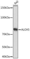
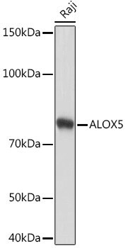
- Western blot analysis of ALOX5 in extracts from Raji cells. Samples were incubated with ALOX5 Monoclonal antibody (Product # MA5-38050) using a dilution of 1:1,000, followed by HRP Goat Anti-Rabbit IgG (H+L) at a dilution of 1:10,000. Lysates/proteins: 25 µg per lane. Blocking buffer: 3% nonfat dry milk in TBST. Detection: ECL Basic Kit. Exposure time: 10s.
- Submitted by
- Invitrogen Antibodies (provider)
- Main image
- Experimental details
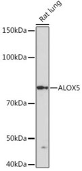
- Western blot analysis of ALOX5 in extracts of Rat lung. Samples were incubated with ALOX5 Monoclonal antibody (Product # MA5-38050) using a dilution of 1:1,000, followed by HRP Goat Anti-Rabbit IgG (H+L) at a dilution of 1:10,000. Lysates/proteins: 25 µg per lane. Blocking buffer: 3% nonfat dry milk in TBST. Detection: ECL Basic Kit. Exposure time: 3 min.

Supportive validation
- Submitted by
- Invitrogen Antibodies (provider)
- Main image
- Experimental details
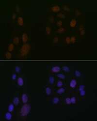
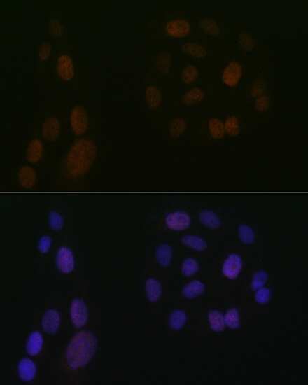
- Immunocytochemistry/Immunofluorescence analysis of ALOX5 in U-2 OS cells using ALOX5 Recombinant Monoclonal Antibody (Product # MA5-38050) at a dilution of 1:100. Blue: DAPI for nuclear staining.
- Submitted by
- Invitrogen Antibodies (provider)
- Main image
- Experimental details
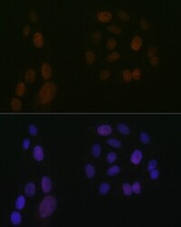
- Immunocytochemistry/Immunofluorescence analysis of ALOX5 in U-2 OS cells using ALOX5 Recombinant Monoclonal Antibody (Product # MA5-38050) at a dilution of 1:100. Blue: DAPI for nuclear staining.

- Submitted by
- Invitrogen Antibodies (provider)
- Main image

- Experimental details
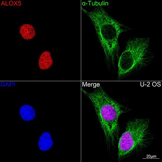
- Confocal imaging (Immunocytochemistry) of ALOX5 in U-2 OS cells. Samples were incubated with ALOX5 Monoclonal antibody (Product # MA5-38050) using a dilution of 1:100 (Red). The cells were counterstained with alpha Tubulin Mouse mAb (dilution 1:400) (Green). DAPI was used for nuclear staining (blue). Objective: 100x.
Supportive validation
- Submitted by
- Invitrogen Antibodies (provider)
- Main image
- Experimental details
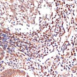
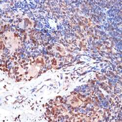
- Immunohistochemistry analysis of ALOX5 in paraffin-embedded human appendix. Samples were incubated with ALOX5 Monoclonal antibody (Product # MA5-38050) using a dilution of 1:100 (40x lens). Perform microwave antigen retrieval with 10 mM Tris/EDTA buffer pH 9.0 before commencing with IHC staining protocol.

- Submitted by
- Invitrogen Antibodies (provider)
- Main image
- Experimental details
- Immunohistochemistry analysis of ALOX5 in paraffin-embedded mouse spleen. Samples were incubated with ALOX5 Monoclonal antibody (Product # MA5-38050) using a dilution of 1:100 (40x lens). Perform microwave antigen retrieval with 10 mM Tris/EDTA buffer pH 9.0 before commencing with IHC staining protocol.
