Explore
 Validate
 Learn
LearnMA1-513
antibody from Invitrogen Antibodies
Targeting: AHR
bHLHe76
 Western blot
 Immunocytochemistry
 Immunoprecipitation Immunohistochemistry Flow cytometry Gel shift Chromatin Immunoprecipitation Other assay
Antibody data
- Antibody Data
- Antigen structure
- References [24]
- Comments [0]
- Validations
- Immunocytochemistry [6]
- Flow cytometry [5]
- Other assay [12]
Submit
Validation data
Reference
Comment
Report error
- Product number
- MA1-513 - Provider product page

- Provider
- Invitrogen Antibodies
- Product name
- AHR Monoclonal Antibody (RPT9)
- Antibody type
- Monoclonal
- Antigen
- Synthetic peptide
- Description
- MA1-513 detects the aryl hydrocarbon receptor (AhR) from human, mouse, and rat cells. MA1-513 has been successfully used in immunoprecipitation, chromatin immunoprecipitation, immunocytochemistry, immunohistochemistry, immunofluorescence, and gel shift procedures. This antibody specifically immunoprecipitates a single ~95 kDa protein representing AhR from Hepa 1 cytosol. Although MA1-513 can be used in Western blotting procedures, MA1-514 (clone RPT1) is recommended for this procedure. The MA1-513 immunogen is a synthetic peptide corresponding to residues R(12) K R R K P(17) V(22) K P I P A E G I K(31) of mouse AhR.
- Reactivity
- Human, Mouse, Rat
- Host
- Mouse
- Isotype
- IgG
- Antibody clone number
- RPT9
- Vial size
- 100 μL
- Concentration
- 1 mg/mL
- Storage
- -20°C, Avoid Freeze/Thaw Cycles
Submitted references Anakinra restores cellular proteostasis by coupling mitochondrial redox balance to autophagy.
LINC00665 knockdown confers sensitivity in irradiated non-small cell lung cancer cells through the miR-582-5p/UCHL3/AhR axis.
Bioengineering of Genetically Encoded Gene Promoter Repressed by the Flavonoid Apigenin for Constructing Intracellular Sensor for Molecular Events.
AhR activation attenuates calcium oxalate nephrocalcinosis by diminishing M1 macrophage polarization and promoting M2 macrophage polarization.
Immunoregulatory effects of indole-3-carbinol on monocyte-derived macrophages in systemic lupus erythematosus: A crucial role for aryl hydrocarbon receptor.
Quaking Deficiency Amplifies Inflammation in Experimental Endotoxemia via the Aryl Hydrocarbon Receptor/Signal Transducer and Activator of Transcription 1-NF-κB Pathway.
The activation mechanism of the aryl hydrocarbon receptor (AhR) by molecular chaperone HSP90.
Tobacco smoke-induced skin pigmentation is mediated by the aryl hydrocarbon receptor.
Effect of transient TCDD exposure on immortalized human trophoblast-derived cell lines.
A role of the aryl hydrocarbon receptor in attenuation of colitis.
A 5'-region polymorphism modulates promoter activity of the tumor suppressor gene MFSD2A.
Aryl hydrocarbon receptor-mediated up-regulation of ATP-driven xenobiotic efflux transporters at the blood-brain barrier.
Aryl hydrocarbon receptor functions as a potent coactivator of E2F1-dependent trascription activity.
Ube2l3 gene expression is modulated by activation of the aryl hydrocarbon receptor: implications for p53 ubiquitination.
2,3,7,8-tetrachlorodibenzo-p-dioxin impairs an insulin signaling pathway through the induction of tumor necrosis factor-alpha in adipocytes.
Cytochromes P4501 (CYP1): catalytic activities and inducibility by diesel exhaust particle extract and benzo[a]pyrene in intact human lung ex vivo.
Aryl hydrocarbon receptor is a transcriptional activator of the human breast cancer resistance protein (BCRP/ABCG2).
Role of GAC63 in transcriptional activation mediated by the aryl hydrocarbon receptor.
2,3,7,8-Tetrachlorodibenzo-p-dioxin induces insulin-like growth factor binding protein-1 gene expression and counteracts the negative effect of insulin.
Role of the coiled-coil coactivator (CoCoA) in aryl hydrocarbon receptor-mediated transcription.
Dietary polyphenols increase paraoxonase 1 gene expression by an aryl hydrocarbon receptor-dependent mechanism.
A new southwestern chemistry-based ELISA for detection of aryl hydrocarbon receptor transformation: application to the screening of its receptor agonists and antagonists.
7-ketocholesterol is an endogenous modulator for the arylhydrocarbon receptor.
Characterization of the AhR-hsp90-XAP2 core complex and the role of the immunophilin-related protein XAP2 in AhR stabilization.
van de Veerdonk FL, Renga G, Pariano M, Bellet MM, Servillo G, Fallarino F, De Luca A, Iannitti RG, Piobbico D, Gargaro M, Manni G, D'Onofrio F, Stincardini C, Sforna L, Borghi M, Castelli M, Pieroni S, Oikonomou V, Villella VR, Puccetti M, Giovagnoli S, Galarini R, Barola C, Maiuri L, Della Fazia MA, Cellini B, Talesa VN, Dinarello CA, Costantini C, Romani L
The Journal of clinical investigation 2022 Jan 18;132(2)
The Journal of clinical investigation 2022 Jan 18;132(2)
LINC00665 knockdown confers sensitivity in irradiated non-small cell lung cancer cells through the miR-582-5p/UCHL3/AhR axis.
Xu LM, Yuan YJ, Yu H, Wang S, Wang P
Journal of translational medicine 2022 Aug 2;20(1):350
Journal of translational medicine 2022 Aug 2;20(1):350
Bioengineering of Genetically Encoded Gene Promoter Repressed by the Flavonoid Apigenin for Constructing Intracellular Sensor for Molecular Events.
Desmet NM, Dhusia K, Qi W, Doseff AI, Bhattacharya S, Gilad AA
Biosensors 2021 Apr 28;11(5)
Biosensors 2021 Apr 28;11(5)
AhR activation attenuates calcium oxalate nephrocalcinosis by diminishing M1 macrophage polarization and promoting M2 macrophage polarization.
Yang X, Liu H, Ye T, Duan C, Lv P, Wu X, Liu J, Jiang K, Lu H, Yang H, Xia D, Peng E, Chen Z, Tang K, Ye Z
Theranostics 2020;10(26):12011-12025
Theranostics 2020;10(26):12011-12025
Immunoregulatory effects of indole-3-carbinol on monocyte-derived macrophages in systemic lupus erythematosus: A crucial role for aryl hydrocarbon receptor.
Mohammadi S, Memarian A, Sedighi S, Behnampour N, Yazdani Y
Autoimmunity 2018 Aug;51(5):199-209
Autoimmunity 2018 Aug;51(5):199-209
Quaking Deficiency Amplifies Inflammation in Experimental Endotoxemia via the Aryl Hydrocarbon Receptor/Signal Transducer and Activator of Transcription 1-NF-κB Pathway.
Wang L, Zhai DS, Ruan BJ, Xu CM, Ye ZC, Lu HY, Jiang YH, Wang ZY, Xiang A, Yang Y, Yuan JL, Lu ZF
Frontiers in immunology 2017;8:1754
Frontiers in immunology 2017;8:1754
The activation mechanism of the aryl hydrocarbon receptor (AhR) by molecular chaperone HSP90.
Tsuji N, Fukuda K, Nagata Y, Okada H, Haga A, Hatakeyama S, Yoshida S, Okamoto T, Hosaka M, Sekine K, Ohtaka K, Yamamoto S, Otaka M, Grave E, Itoh H
FEBS open bio 2014;4:796-803
FEBS open bio 2014;4:796-803
Tobacco smoke-induced skin pigmentation is mediated by the aryl hydrocarbon receptor.
Nakamura M, Ueda Y, Hayashi M, Kato H, Furuhashi T, Morita A
Experimental dermatology 2013 Aug;22(8):556-8
Experimental dermatology 2013 Aug;22(8):556-8
Effect of transient TCDD exposure on immortalized human trophoblast-derived cell lines.
Fukushima K, Tsukimori K, Li D, Takao T, Morokuma S, Kato K, Seki H, Takeda S, Matsumura S, Wake N
Human & experimental toxicology 2012 Jun;31(6):550-6
Human & experimental toxicology 2012 Jun;31(6):550-6
A role of the aryl hydrocarbon receptor in attenuation of colitis.
Furumatsu K, Nishiumi S, Kawano Y, Ooi M, Yoshie T, Shiomi Y, Kutsumi H, Ashida H, Fujii-Kuriyama Y, Azuma T, Yoshida M
Digestive diseases and sciences 2011 Sep;56(9):2532-44
Digestive diseases and sciences 2011 Sep;56(9):2532-44
A 5'-region polymorphism modulates promoter activity of the tumor suppressor gene MFSD2A.
Colombo F, Falvella FS, Galvan A, Frullanti E, Kunitoh H, Ushijima T, Dragani TA
Molecular cancer 2011 Jul 7;10:81
Molecular cancer 2011 Jul 7;10:81
Aryl hydrocarbon receptor-mediated up-regulation of ATP-driven xenobiotic efflux transporters at the blood-brain barrier.
Wang X, Hawkins BT, Miller DS
FASEB journal : official publication of the Federation of American Societies for Experimental Biology 2011 Feb;25(2):644-52
FASEB journal : official publication of the Federation of American Societies for Experimental Biology 2011 Feb;25(2):644-52
Aryl hydrocarbon receptor functions as a potent coactivator of E2F1-dependent trascription activity.
Watabe Y, Nazuka N, Tezuka M, Shimba S
Biological & pharmaceutical bulletin 2010;33(3):389-97
Biological & pharmaceutical bulletin 2010;33(3):389-97
Ube2l3 gene expression is modulated by activation of the aryl hydrocarbon receptor: implications for p53 ubiquitination.
Reyes-Hernández OD, Mejía-García A, Sánchez-Ocampo EM, Cabañas-Cortés MA, Ramírez P, Chávez-González L, Gonzalez FJ, Elizondo G
Biochemical pharmacology 2010 Sep 15;80(6):932-40
Biochemical pharmacology 2010 Sep 15;80(6):932-40
2,3,7,8-tetrachlorodibenzo-p-dioxin impairs an insulin signaling pathway through the induction of tumor necrosis factor-alpha in adipocytes.
Nishiumi S, Yoshida M, Azuma T, Yoshida K, Ashida H
Toxicological sciences : an official journal of the Society of Toxicology 2010 Jun;115(2):482-91
Toxicological sciences : an official journal of the Society of Toxicology 2010 Jun;115(2):482-91
Cytochromes P4501 (CYP1): catalytic activities and inducibility by diesel exhaust particle extract and benzo[a]pyrene in intact human lung ex vivo.
Iba MM, Shin M, Caccavale RJ
Toxicology 2010 Jun 29;273(1-3):35-44
Toxicology 2010 Jun 29;273(1-3):35-44
Aryl hydrocarbon receptor is a transcriptional activator of the human breast cancer resistance protein (BCRP/ABCG2).
Tan KP, Wang B, Yang M, Boutros PC, Macaulay J, Xu H, Chuang AI, Kosuge K, Yamamoto M, Takahashi S, Wu AM, Ross DD, Harper PA, Ito S
Molecular pharmacology 2010 Aug;78(2):175-85
Molecular pharmacology 2010 Aug;78(2):175-85
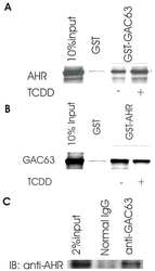
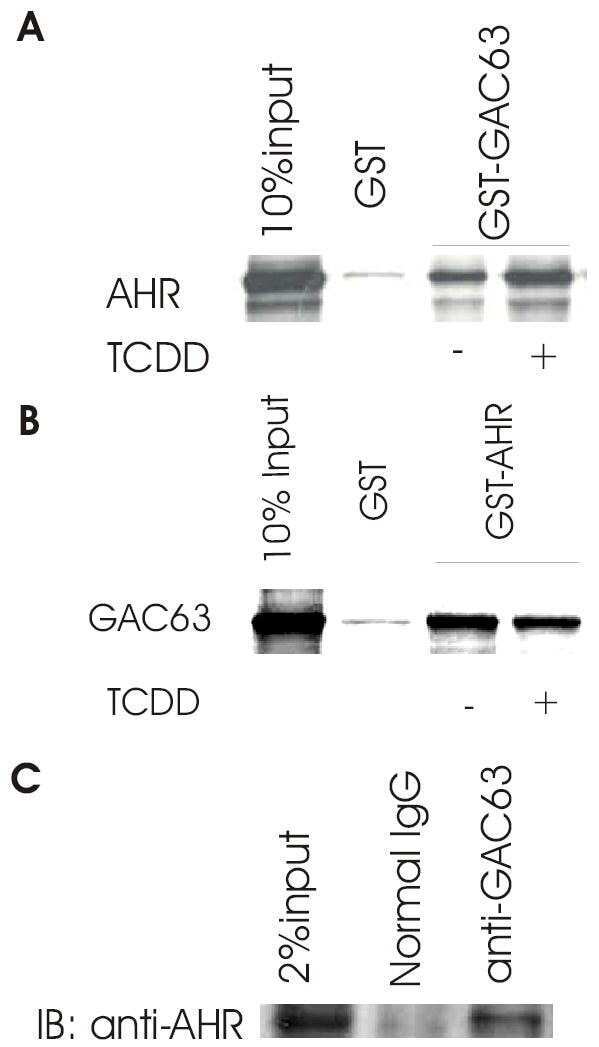
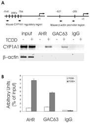
Role of GAC63 in transcriptional activation mediated by the aryl hydrocarbon receptor.
Chen YH, Beischlag TV, Kim JH, Perdew GH, Stallcup MR
The Journal of biological chemistry 2006 May 5;281(18):12242-7
The Journal of biological chemistry 2006 May 5;281(18):12242-7
2,3,7,8-Tetrachlorodibenzo-p-dioxin induces insulin-like growth factor binding protein-1 gene expression and counteracts the negative effect of insulin.
Marchand A, Tomkiewicz C, Marchandeau JP, Boitier E, Barouki R, Garlatti M
Molecular pharmacology 2005 Feb;67(2):444-52
Molecular pharmacology 2005 Feb;67(2):444-52
Role of the coiled-coil coactivator (CoCoA) in aryl hydrocarbon receptor-mediated transcription.
Kim JH, Stallcup MR
The Journal of biological chemistry 2004 Nov 26;279(48):49842-8
The Journal of biological chemistry 2004 Nov 26;279(48):49842-8
Dietary polyphenols increase paraoxonase 1 gene expression by an aryl hydrocarbon receptor-dependent mechanism.
Gouédard C, Barouki R, Morel Y
Molecular and cellular biology 2004 Jun;24(12):5209-22
Molecular and cellular biology 2004 Jun;24(12):5209-22
A new southwestern chemistry-based ELISA for detection of aryl hydrocarbon receptor transformation: application to the screening of its receptor agonists and antagonists.
Fukuda I, Nishiumi S, Yabushita Y, Mukai R, Kodoi R, Hashizume K, Mizuno M, Hatanaka Y, Ashida H
Journal of immunological methods 2004 Apr;287(1-2):187-201
Journal of immunological methods 2004 Apr;287(1-2):187-201
7-ketocholesterol is an endogenous modulator for the arylhydrocarbon receptor.
Savouret JF, Antenos M, Quesne M, Xu J, Milgrom E, Casper RF
The Journal of biological chemistry 2001 Feb 2;276(5):3054-9
The Journal of biological chemistry 2001 Feb 2;276(5):3054-9
Characterization of the AhR-hsp90-XAP2 core complex and the role of the immunophilin-related protein XAP2 in AhR stabilization.
Meyer BK, Perdew GH
Biochemistry 1999 Jul 13;38(28):8907-17
Biochemistry 1999 Jul 13;38(28):8907-17
No comments: Submit comment
Supportive validation
- Submitted by
- Invitrogen Antibodies (provider)
- Main image

- Experimental details
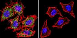
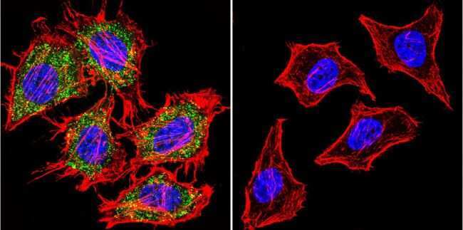
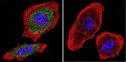
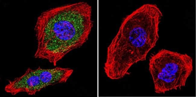
- Immunofluorescent analysis of Aryl Hydrocarbon Receptor using Aryl Hydrocarbon Receptor Monoclonal Antibody (RPT9) (Product # MA1-513) shows staining in A2058 Cells. Aryl Hydrocarbon Receptor (green), F-Actin staining with Phalloidin (red) and nuclei with DAPI (blue) is shown. Cells were grown on chamber slides and fixed with formaldehyde prior to staining. Cells were probed without (control) or with an antibody recognizing Aryl Hydrocarbon Receptor (Product # MA1-513) at a dilution of 1:20 over night at 4 °C, washed with PBS and incubated with a DyLight-488 conjugated secondary antibody (Product # 35552 for GAR, Product # 35503 for GAM). Images were taken at 60X magnification.

- Submitted by
- Invitrogen Antibodies (provider)
- Main image
- Experimental details
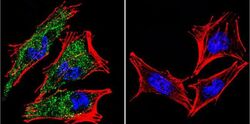
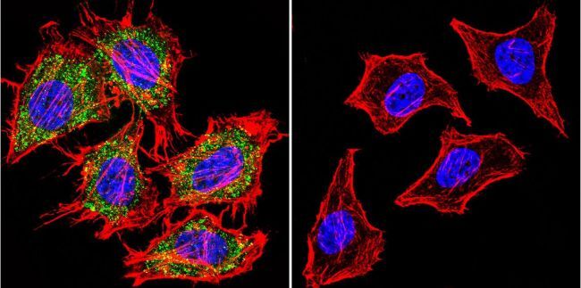
- Immunofluorescent analysis of Aryl Hydrocarbon Receptor using Aryl Hydrocarbon Receptor Monoclonal Antibody (RPT9) (Product # MA1-513) shows staining in Hela Cells. Aryl Hydrocarbon Receptor (green), F-Actin staining with Phalloidin (red) and nuclei with DAPI (blue) is shown. Cells were grown on chamber slides and fixed with formaldehyde prior to staining. Cells were probed without (control) or with an antibody recognizing Aryl Hydrocarbon Receptor (Product # MA1-513) at a dilution of 1:20 over night at 4 °C, washed with PBS and incubated with a DyLight-488 conjugated secondary antibody (Product # 35552 for GAR, Product # 35503 for GAM). Images were taken at 60X magnification.
- Submitted by
- Invitrogen Antibodies (provider)
- Main image
- Experimental details
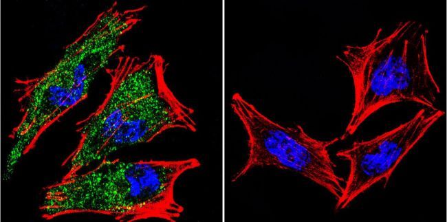
- Immunofluorescent analysis of Aryl Hydrocarbon Receptor using Aryl Hydrocarbon Receptor Monoclonal Antibody (RPT9) (Product # MA1-513) shows staining in U251 Cells. Aryl Hydrocarbon Receptor (green), F-Actin staining with Phalloidin (red) and nuclei with DAPI (blue) is shown. Cells were grown on chamber slides and fixed with formaldehyde prior to staining. Cells were probed without (control) or with an antibody recognizing Aryl Hydrocarbon Receptor (Product # MA1-513) at a dilution of 1:20 over night at 4 °C, washed with PBS and incubated with a DyLight-488 conjugated secondary antibody (Product # 35552 for GAR, Product # 35503 for GAM). Images were taken at 60X magnification.
- Submitted by
- Invitrogen Antibodies (provider)
- Main image
- Experimental details
- Immunofluorescent analysis of Aryl Hydrocarbon Receptor using Aryl Hydrocarbon Receptor Monoclonal Antibody (RPT9) (Product # MA1-513) shows staining in A2058 Cells. Aryl Hydrocarbon Receptor (green), F-Actin staining with Phalloidin (red) and nuclei with DAPI (blue) is shown. Cells were grown on chamber slides and fixed with formaldehyde prior to staining. Cells were probed without (control) or with an antibody recognizing Aryl Hydrocarbon Receptor (Product # MA1-513) at a dilution of 1:20 over night at 4 °C, washed with PBS and incubated with a DyLight-488 conjugated secondary antibody (Product # 35552 for GAR, Product # 35503 for GAM). Images were taken at 60X magnification.
- Submitted by
- Invitrogen Antibodies (provider)
- Main image
- Experimental details
- Immunofluorescent analysis of Aryl Hydrocarbon Receptor using Aryl Hydrocarbon Receptor Monoclonal Antibody (RPT9) (Product # MA1-513) shows staining in Hela Cells. Aryl Hydrocarbon Receptor (green), F-Actin staining with Phalloidin (red) and nuclei with DAPI (blue) is shown. Cells were grown on chamber slides and fixed with formaldehyde prior to staining. Cells were probed without (control) or with an antibody recognizing Aryl Hydrocarbon Receptor (Product # MA1-513) at a dilution of 1:20 over night at 4 °C, washed with PBS and incubated with a DyLight-488 conjugated secondary antibody (Product # 35552 for GAR, Product # 35503 for GAM). Images were taken at 60X magnification.
- Submitted by
- Invitrogen Antibodies (provider)
- Main image

- Experimental details
- Immunofluorescent analysis of Aryl Hydrocarbon Receptor using Aryl Hydrocarbon Receptor Monoclonal Antibody (RPT9) (Product # MA1-513) shows staining in U251 Cells. Aryl Hydrocarbon Receptor (green), F-Actin staining with Phalloidin (red) and nuclei with DAPI (blue) is shown. Cells were grown on chamber slides and fixed with formaldehyde prior to staining. Cells were probed without (control) or with an antibody recognizing Aryl Hydrocarbon Receptor (Product # MA1-513) at a dilution of 1:20 over night at 4 °C, washed with PBS and incubated with a DyLight-488 conjugated secondary antibody (Product # 35552 for GAR, Product # 35503 for GAM). Images were taken at 60X magnification.

Supportive validation
- Submitted by
- Invitrogen Antibodies (provider)
- Main image
- Experimental details
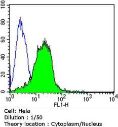
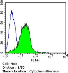
- Flow cytometry analysis of Aryl Hydrocarbon Receptor in Hela cells compared to an isotype control (blue). Cells were harvested, adjusted to a concentration of 1-5x10^6 cells/mL, fixed with 2% paraformaldehyde and washed with PBS. Cells were penetrated by dropping the supernatant, adding 90% methanol and incubated for 10 minutes at room temperature. Follwing penetration, cells were blocked with a 2% solution of BSA-PBS for 30 min at room temperature and incubated with a Aryl Hydrocarbon Receptor monoclonal antibody (Product # MA1-513) at a dilution of 1:50 for 60 min at room temperature. Cells were then incubated for 40 min at room temperature in the dark using a Dylight 488-conjugated goat anti-mouse IgG (H+L) secondary antibody and re-suspended in PBS for FACS analysis.

- Submitted by
- Invitrogen Antibodies (provider)
- Main image

- Experimental details
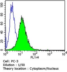
- Flow cytometry analysis of Aryl Hydrocarbon Receptor in PC-3 cells compared to an isotype control (blue). Cells were harvested, adjusted to a concentration of 1-5x10^6 cells/mL, fixed with 2% paraformaldehyde and washed with PBS. Cells were penetrated by dropping the supernatant, adding 90% methanol and incubated for 10 minutes at room temperature. Follwing penetration, cells were blocked with a 2% solution of BSA-PBS for 30 min at room temperature and incubated with a Aryl Hydrocarbon Receptor monoclonal antibody (Product # MA1-513) at a dilution of 1:50 for 60 min at room temperature. Cells were then incubated for 40 min at room temperature in the dark using a Dylight 488-conjugated goat anti-mouse IgG (H+L) secondary antibody and re-suspended in PBS for FACS analysis.

- Submitted by
- Invitrogen Antibodies (provider)
- Main image
- Experimental details
- Flow cytometry analysis of Aryl Hydrocarbon Receptor in Hela cells compared to an isotype control (blue). Cells were harvested, adjusted to a concentration of 1-5x10^6 cells/mL, fixed with 2% paraformaldehyde and washed with PBS. Cells were penetrated by dropping the supernatant, adding 90% methanol and incubated for 10 minutes at room temperature. Follwing penetration, cells were blocked with a 2% solution of BSA-PBS for 30 min at room temperature and incubated with a Aryl Hydrocarbon Receptor monoclonal antibody (Product # MA1-513) at a dilution of 1:50 for 60 min at room temperature. Cells were then incubated for 40 min at room temperature in the dark using a Dylight 488-conjugated goat anti-mouse IgG (H+L) secondary antibody and re-suspended in PBS for FACS analysis.

- Submitted by
- Invitrogen Antibodies (provider)
- Main image
- Experimental details
- Flow cytometry analysis of Aryl Hydrocarbon Receptor in PC-3 cells compared to an isotype control (blue). Cells were harvested, adjusted to a concentration of 1-5x10^6 cells/mL, fixed with 2% paraformaldehyde and washed with PBS. Cells were penetrated by dropping the supernatant, adding 90% methanol and incubated for 10 minutes at room temperature. Follwing penetration, cells were blocked with a 2% solution of BSA-PBS for 30 min at room temperature and incubated with a Aryl Hydrocarbon Receptor monoclonal antibody (Product # MA1-513) at a dilution of 1:50 for 60 min at room temperature. Cells were then incubated for 40 min at room temperature in the dark using a Dylight 488-conjugated goat anti-mouse IgG (H+L) secondary antibody and re-suspended in PBS for FACS analysis.

- Submitted by
- Invitrogen Antibodies (provider)
- Main image
- Experimental details
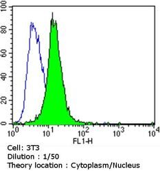
- Flow cytometry analysis of Aryl Hydrocarbon Receptor in NIH/3T3 cells compared to an isotype control (blue). Cells were harvested, adjusted to a concentration of 1-5x10^6 cells/mL, fixed with 2% paraformaldehyde and washed with PBS. Cells were penetrated by dropping the supernatant, adding 90% methanol and incubated for 10 minutes at room temperature. Follwing penetration, cells were blocked with a 2% solution of BSA-PBS for 30 min at room temperature and incubated with a Aryl Hydrocarbon Receptor monoclonal antibody (Product # MA1-513) at a dilution of 1:50 for 60 min at room temperature. Cells were then incubated for 40 min at room temperature in the dark using a Dylight 488-conjugated goat anti-mouse IgG (H+L) secondary antibody and re-suspended in PBS for FACS analysis.

Supportive validation
- Submitted by
- Invitrogen Antibodies (provider)
- Main image
- Experimental details
- NULL
- Submitted by
- Invitrogen Antibodies (provider)
- Main image
- Experimental details
- NULL

- Submitted by
- Invitrogen Antibodies (provider)
- Main image
- Experimental details
- NULL

- Submitted by
- Invitrogen Antibodies (provider)
- Main image
- Experimental details
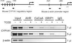
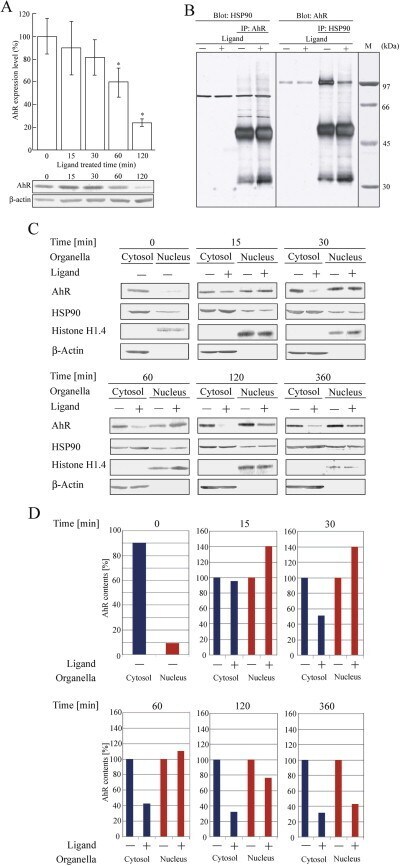
- Fig. 5 Total amounts of endogenous AhR in the cytosol and nucleus of HeLa cells. (A) HeLa cells were treated with 0.05% DMSO (-) or 1 muM beta-NF (+) for 0, 15, 30, 60, and 120 min at 37 degC. The lysate was collected and centrifuged for 60 min at 105,000 g , then the supernatant was collected. AhR in the cytosol were analyzed by immnoblotting with an anti-AhR antibody or an anti- beta-actin antibody ( n = 4, * p < 0.05). The AhR/beta-actin ratio was quantified using Image J software (Drop of Wisdom). (B) HeLa cells were treated with 0.05% DMSO (-) or 3 muM beta-NF (+) for 30 min at 37 degC. The lysate was collected and centrifuged, then the supernatant was collected. Immunoprecipitation was performed using an anti-Ahr antibody or anti-HSP90 antibody followed by immunoblotting with an anti-HSP90 antibody or an anti-AhR antibody. In panel, the HeLa cell lysate (left two lanes) or immunoprecipitated samples (right two lanes) were separated by SDS-PAGE followed by immnoblotting. The subunit molecular weight of HSP90 and AhR are 90 and 110 kDa, respectively. The 50- and 32-kDa protein bands were the IgG heavy- and light chain, respectively. M, molecular marker proteins. C, HeLa cells were treated with 0.05% DMSO (-) or 1 muM beta-NF (+) for 0, 15, 30, 60, 120, and 360 min at 37 degC. The lysate was collected and centrifuged for 60 min at 105,000 g , then the supernatant was collected as a cytosol fraction. Or the lysate in 2.2-M sucrose solution was centrifuged for 60 min at 30,0
- Submitted by
- Invitrogen Antibodies (provider)
- Main image
- Experimental details
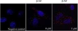
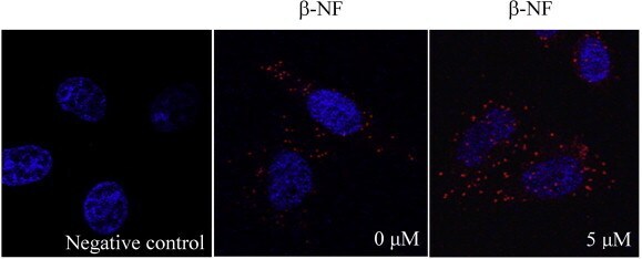
- Fig. 6 beta-NF induced AhR and HSP90 nuclear translocation in HeLa cells. HeLa cells were treated with beta-NF (5 muM) or DMSO (0.1%) for 120 min. Complex formation between HSP90 and AhR was measured by in situ PLA using rabbit anti-AhR antibodies, mouse anti-HSP90 antibodies, and corresponding reagents. In situ PLA is indicated by red signals. Blue staining indicates DAPI staining of cell nuclei. Control was stained with non-immune rabbit- and mouse serum. (For interpretation of the references to color in this figure legend, the reader is referred to the web version of this article.)
- Submitted by
- Invitrogen Antibodies (provider)
- Main image

- Experimental details
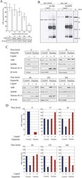
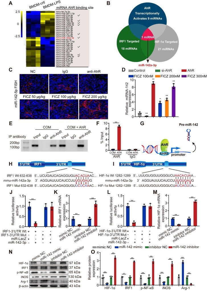
- Figure 4 AhR transcriptionally activates miR-142a to inhibit IRF1 and HIF-1alpha expression. (A) The top 30 miRNAs in BMDMs that are regulated by LPS are arranged in a miRNA array heatmap. In addition, miRNAs predicted to be under the transcriptional control of AhR (according to analysis with the JASPAR database) are noted. (B) Venn diagram analyses were performed to identify miRNAs that can both target IRF1 and HIF-1alpha and that are under the transcriptional control of AhR. (C) Renal expression of mmu-miR-142a-3p in mice (n = 6) with CaOx nephrocalcinosis following treatment with an AhR neutralizing antibody or FICZ treatment was assessed via FISH (200x; scale bar: 20 um). (D) qRT-PCR was performed to measure mmu-miR-142a-3p expression in BMDMs using U6 RNA as a normalization control. (E, F) ChIP assays and ChIP qPCR analysis showed that AhR bound to the miR-142a promoter in BMDMs treated with the AhR overexpression plasmid. (G) A schematic model showed that AhR directly binds to the miR-142a promoter and activates its transcription. (H, I) WT and mutated miR-142a targeting sequences in the IRF1 and HIF-1alpha 3'-UTR regions that were used to construct luciferase reporters, with reporters bearing these IRF1 (J) or HIF-1alpha (L) 3'-UTR sequences co-transfected along with miR-142a mimic (100 nM). IRF1 (K) and HIF-1alpha (M) mRNA levels were detected via qRT-PCR in BMDMs following miR-142a mimic or inhibitor transfection. Western blotting (N, O) analysis enabled the detectio
- Submitted by
- Invitrogen Antibodies (provider)
- Main image
- Experimental details
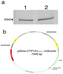
- Figure 2 ( a ) Western blot analysis of AHR protein expression in HEK293FT (1) and HeLa (2). A band represents the AHR was observed at 95kD as expected. ( b ) Vector map of pGlow-CYP1A1 prom ::mScarlet plasmid.

- Submitted by
- Invitrogen Antibodies (provider)
- Main image
- Experimental details
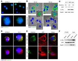
- Figure 4 Anakinra colocalizes with AhR in Il1r1 -/- cells. ( A ) Immunofluorescence imaging of cellular localization of FITC-anakinra in alveolar macrophages, isolated from naive C57BL/6 or Il1r1 -/- mice, primed with 100 ng/mL LPS for 2 hours at 37degC, and exposed to 10 mug/mL FITC-anakinra for 60 minutes. ( B ) Immunofluorescence analysis of FITC-anakinra in Il1r1 -/- cells exposed to FITC-anakinra at 30 and 60 minutes in the presence of 5 muM cytochalasin D. Insets, percentage positive cells. ( C ) Il1r1 -/- MEF cells were treated with 10 mug/mL anakinra for 0, 2, and 4 hours and assessed for AhR and IL1Ra expression by immunoblotting with specific antibodies. ( D ) Immunofluorescence imaging of alveolar macrophages from naive C57BL/6 and Il1r1 -/- mice primed with 100 ng/mL LPS for 2 hours at 37degC, exposed to 10 mug/mL FITC-anakinra for 60 minutes, and stained with anti-AhR antibody. ( E ) Immunofluorescence imaging of MEF cells stained with anti-AhR antibody and DAPI and exposed to 10 mug/mL FITC-anakinra for 60 minutes. ( F ) Il1r1 -/- MEF cells were treated with 10 mug/mL anakinra, lysed, immunoprecipitated with anti-AhR antibody, and assessed for IL1Ra and AhR expression by immunoblotting with specific antibodies. None, untreated cells. Representative images, acquired with a fluorescent microscope (BX51), and immunoblots from 2 independent experiments are shown. DAPI was used to detect nuclei. Sections were examined using a Zeiss Axio Observer Z1.

- Submitted by
- Invitrogen Antibodies (provider)
- Main image
- Experimental details
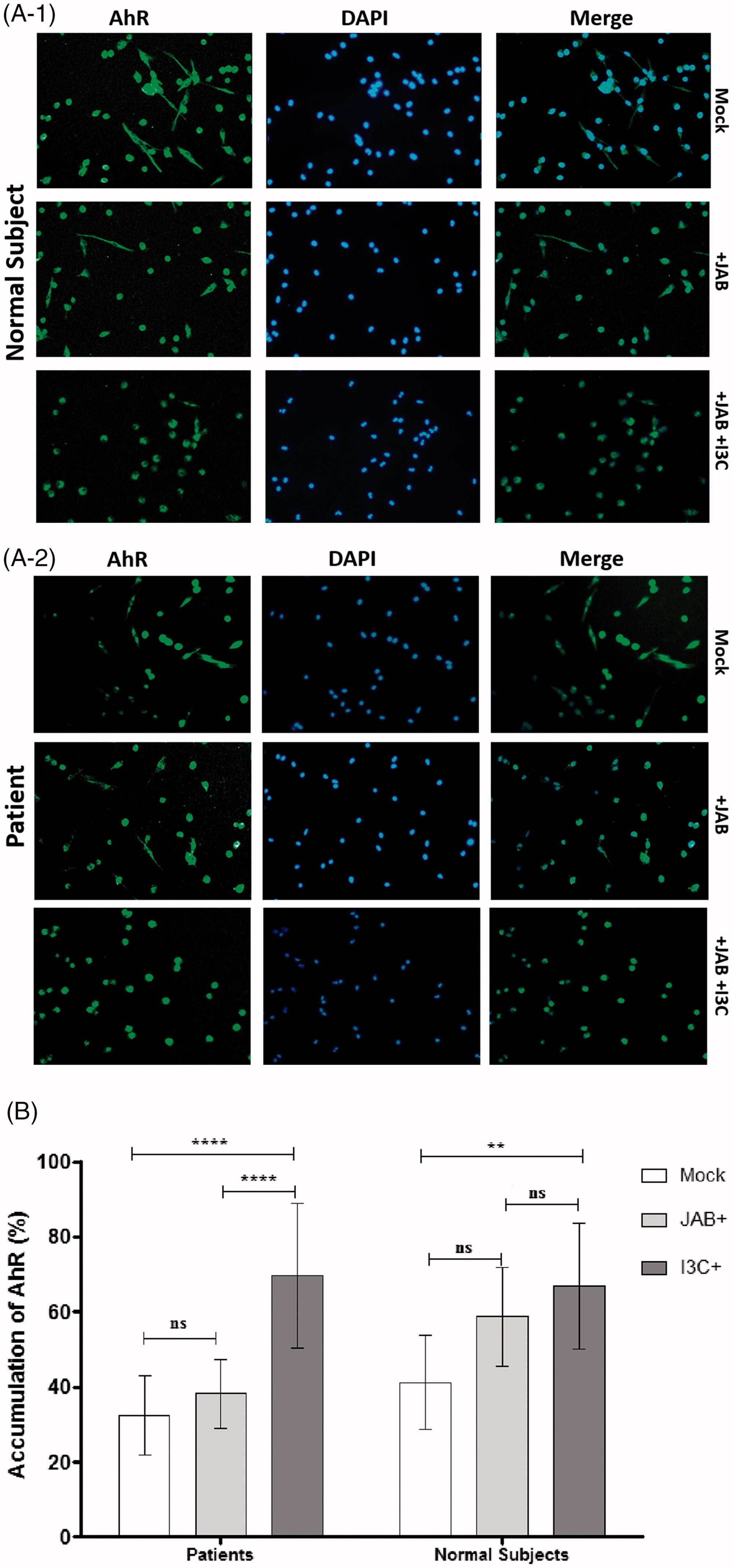
- Figure 5 Anakinra activates a xenobiotic sensing pathway via IDO1. ( A ) H1L1 cells were treated with different doses of kynurenine and anakinra for 6 hours and assessed for luciferase assay. ( B ) Representative immunoblots of IL1RA, HSP90, AIP, and AhR and ( C ) ARNT and AhR in cell lysates in which AhR was immunoprecipitated from Il1r1 -/- MEF cells treated with 10 mug/mL anakinra or 10 muM FICZ. In B and C , data are representative of 1 out of 2 independent experiments and the relative densitometric analysis are reported. ( D and E ) Il1r1 -/- MEF cells were treated with 10 mug/mL anakinra for 2 and 8 hours and analyzed for gene expression by a custom QuantiGene plex gene expression assay. Fold changes are reported as heatmap for 2 and 8 hours ( D ) and histograms for 8 hours ( E ) (data are representative of 1 out of 2 independent experiments). ( F ) Il1r1 -/- MEF cells were treated with 10 mug/mL anakinra and analyzed for mRNA expression of selected genes by RT-PCR ( n = 3 independent samples) and protein expression of IDO1 and SOCS3 by immunoblotting (representative experiment). The relative densitometric analysis is reported. ( G ) Il1r1 -/- MEF cells were treated with either 10 mug/mL anakinra for different times or 10 ng/mL IFN-gamma as positive control for 48 hours and assessed for kynurenine production by ELISA. ( H ) Il1r1 -/- MEF cells were treated with 10 mug/mL anakinra in the presence or absence of 10 muM epacadostat and assessed for kynurenine production by

- Submitted by
- Invitrogen Antibodies (provider)
- Main image
- Experimental details
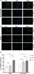
- Figure 1. The cytoplasmic and nuclear localization of AhR in I3C-treated macrophages. The cytoplasmic and nuclear expression of AhR is demonstrated. The nuclear accumulation of AhR was observed upon I3C treatment among MDMs of SLE patients and normal subjects by immunofluorescence staining (A) . Secondary antibody as the isotype control had no detectable fluorescent emission. Blue DAPI-stained nucleus of MDMs were merged with green-coloured AhR-expressing cells (40X magnification). The percentages of AhR accumulation in each combination are quantified (B ). One-way ANOVA with Tukey's post hoc test were used to compare the means of multiple samples. Data of each bar demonstrates means +- SE. p < .05 were considered as statistically significant. * p < .05, ** p < .01, *** p < .001, and **** p < .0001. NS, not statistically significant; Mock, non-treated monocyte-derived macrophages; JAB, Jurkat apoptotic bodies; I3C, indole-3-carbinol.
- Submitted by
- Invitrogen Antibodies (provider)
- Main image
- Experimental details
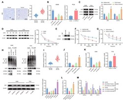
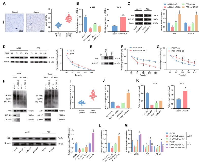
- Fig. 2 UCHL3 maintained the stability of AhR protein through deubiquitination, thereby promoting PD-L1 expression. A , Positive expression of AhR protein in tumor tissues and adjacent normal tissues from NSCLC patients (n = 45) detected by Immunohistochemistry. B , Silencing efficiency of sh-UCHL3 in A549 cells and overexpression efficiency of LV-UCHL3 in PC9 cells detected by qRT-PCR. C , Protein levels of UCHL3 and AhR in A549 cells treated with sh-UCHL3 or in PC9 treated with LV-UCHL3 detected by Western blot analysis. D , Western blot analysis of AhR protein degradation in A549 and PC9 cells following CHX (10 mug/mL) treatment (the left penal) and corresponding statistical analysis of AhR protein stability (the right penal). E , Co-IP detection of exogenous UCHL3 and AhR proteins in HEK293T cells. F-G, AhR protein degradation rate in A549 cells in response to sh-UCHL3 ( F ) or in PC9 cells in response to LV-UCHL3 ( G ) detected by CHX pulse-chase analysis. H , Detection of AhR protein ubiquitination in A549 cells overexpressing UCHL3 or in PC9 cells silencing UCHL3. I , Expression of PD-L1 in tumor tissues and adjacent normal tissues from NSCLC patients (n = 45) detected by qRT-PCR. J , Expression of PD-L1 in A549 cells treated with sh-UCHL3 or in PC9 cells treated with LV-UCHL3 detected by qRT-PCR. K , Silencing efficiency of sh-AhR in A549 cells and overexpression efficiency of LV-AhR in PC9 cells detected by qRT-PCR (the left two panels) and Western blot analysis (the
- Submitted by
- Invitrogen Antibodies (provider)
- Main image

- Experimental details
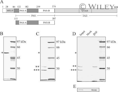
- AhR PAS domain binds to HSP90. (A) Domain structure of human AhR. bHLH, PAS, and TAD indicate basic helix-loop-helix, PER-ARNT-SIM, and transcriptional activation binding domain, respectively. In the present study, we constructed and purified AhR-PAS including PAS-A and PAS-B (115-387). (B) Purified GST-PAS domain was analyzed by SDS-PAGE (9% gel). (C) Digested GST-PAS by factor Xa were separated by SDS-PAGE (9% gel). (D) The factor Xa digested AhR-PAS domains (input) were added to Mock resin (Mock) or beta-naphthoflavone (beta-NF) affinity resin. The bound proteins were separated by SDS-PAGE (9% gel) (D) or by immunoblotting using an anti-AhR antibody (E). In panels B and D, asterisk, double asterisks, and triple asterisks indicate uncut GST-PAS domain, cut PAS domain, and cut GST, respectively.