Explore
 Validate
 Learn
LearnGTX22769
antibody from GeneTex
Targeting: AHR
bHLHe76
 Western blot
 ELISA Immunocytochemistry Immunoprecipitation Immunohistochemistry Flow cytometry Gel shift Chromatin Immunoprecipitation
Antibody data
- Antibody Data
- Antigen structure
- References [0]
- Comments [0]
- Validations
- Western blot [1]
- Immunocytochemistry [3]
- Flow cytometry [3]
Submit
Validation data
Reference
Comment
Report error
- Product number
- GTX22769 - Provider product page

- Provider
- GeneTex
- Proper citation
- GeneTex Cat#GTX22769, RRID:AB_384839
- Product name
- AHR antibody [RPT9]
- Antibody type
- Monoclonal
- Reactivity
- Human, Mouse, Rat
- Host
- Mouse
No comments: Submit comment
Supportive validation
- Submitted by
- GeneTex (provider)
- Main image
- Experimental details
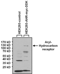
- Western blot analysis of Aryl Hydrocarbon Receptor was performed by loading 40 ug of HEK293 lysate overexpressing Aryl Hydrocarbon Receptor (right lane) or empty vector control (left lane) and 10ul of prestained protein ladder onto a 4-20% Tris-HCl polyacrylamide gel. Proteins were transferred to a PVDF membrane and blocked with 5% BSA/TBST for at least 1 hour. The membrane was probed with an Aryl Hydrocarbon Receptor monoclonal antibody (GTX22769) at a dilution of 1:1000 overnight at 4?C on a rocking platform, washed in TBS-0.1%Tween-20, and probed with a goat anti-mouse IgG-HRP secondary antibody at a dilution of 1:20,000 for 1 hour. Chemiluminescent detection was performed

Supportive validation
- Submitted by
- GeneTex (provider)
- Main image

- Experimental details
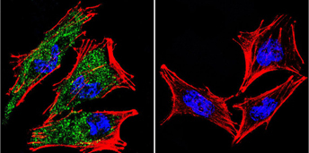
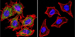
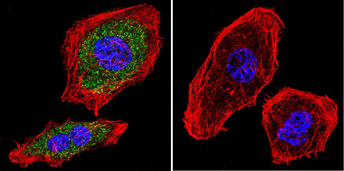
- Immunofluorescent analysis of Aryl Hydrocarbon Receptor using Aryl Hydrocarbon Receptor Monoclonal Antibody (RPT9) (GTX22769) shows staining in A2058 Cells. Aryl Hydrocarbon Receptor (green), F-Actin staining with Phalloidin (red) and nuclei with DAPI (blue) is shown. Cells were grown on chamber slides and fixed with formaldehyde prior to staining. Cells were probed without (control) or with an antibody recognizing Aryl Hydrocarbon Receptor (GTX22769) at a dilution of 1:20 over night at 4 degrees C, washed with PBS and incubated with a DyLight-488 conjugated secondary antibody.
- Submitted by
- GeneTex (provider)
- Main image
- Experimental details
- Immunofluorescent analysis of Aryl Hydrocarbon Receptor using Aryl Hydrocarbon Receptor Monoclonal Antibody (RPT9) (GTX22769) shows staining in Hela Cells. Aryl Hydrocarbon Receptor (green), F-Actin staining with Phalloidin (red) and nuclei with DAPI (blue) is shown. Cells were grown on chamber slides and fixed with formaldehyde prior to staining. Cells were probed without (control) or with an antibody recognizing Aryl Hydrocarbon Receptor (GTX22769) at a dilution of 1:20 over night at 4 degrees C, washed with PBS and incubated with a DyLight-488 conjugated secondary antibody.

- Submitted by
- GeneTex (provider)
- Main image

- Experimental details
- Immunofluorescent analysis of Aryl Hydrocarbon Receptor using Aryl Hydrocarbon Receptor Monoclonal Antibody (RPT9) (GTX22769) shows staining in U251 cells. Aryl Hydrocarbon Receptor (green), F-Actin staining with Phalloidin (red) and nuclei with DAPI (blue) is shown. Cells were grown on chamber slides and fixed with formaldehyde prior to staining. Cells were probed without (control) or with an antibody recognizing Aryl Hydrocarbon Receptor (GTX22769) at a dilution of 1:20 over night at 4 C, washed with PBS and incubated with a DyLight-488 conjugated secondary antibody.
Supportive validation
- Submitted by
- GeneTex (provider)
- Main image

- Experimental details
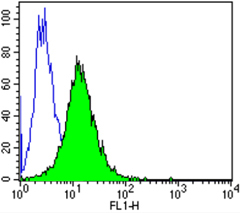
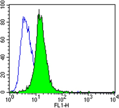
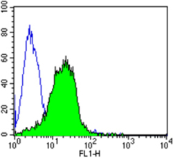
- Flow cytometry analysis of Aryl Hydrocarbon Receptor showing positive staining in the nucleus and cytoplasm of NIH/3T3 cells compared to an isotype control (blue). Cells were harvested, adjusted to a concentration of 1-5x10^6 cells/ml, fixed with 2% paraformaldehyde and washed with PBS. Cells were penetrated by dropping the supernatant, adding 90% methanol and incubated for 10 minutes at room temperature. Follwing penetration, cells were blocked with a 2% solution of BSA-PBS for 30 min at room temperature and incubated with a Aryl Hydrocarbon Receptor monoclonal antibody (GTX22769) at a dilution of 1:50 for 60 min at room temperature. Cells were then incubated for 40 min at room temperature in the dark using a Dylight 488-conjugated goat anti-mouse IgG (H+L) secondary antibody and re-suspended in PBS for FACS analysis.
- Submitted by
- GeneTex (provider)
- Main image
- Experimental details
- Flow cytometry analysis of Aryl Hydrocarbon Receptor showing positive staining in the nucleus and cytoplasm of Hela cells compared to an isotype control (blue). Cells were harvested, adjusted to a concentration of 1-5x10^6 cells/ml, fixed with 2% paraformaldehyde and washed with PBS. Cells were penetrated by dropping the supernatant, adding 90% methanol and incubated for 10 minutes at room temperature. Follwing penetration, cells were blocked with a 2% solution of BSA-PBS for 30 min at room temperature and incubated with a Aryl Hydrocarbon Receptor monoclonal antibody (GTX22769) at a dilution of 1:50 for 60 min at room temperature. Cells were then incubated for 40 min at room temperature in the dark using a Dylight 488-conjugated goat anti-mouse IgG (H+L) secondary antibody and re-suspended in PBS for FACS analysis.

- Submitted by
- GeneTex (provider)
- Main image

- Experimental details
- Flow cytometry analysis of Aryl Hydrocarbon Receptor showing positive staining in the nucleus and cytoplasm of PC-3 cells compared to an isotype control (blue). Cells were harvested, adjusted to a concentration of 1-5x10^6 cells/ml, fixed with 2% paraformaldehyde and washed with PBS. Cells were penetrated by dropping the supernatant, adding 90% methanol and incubated for 10 minutes at room temperature. Follwing penetration, cells were blocked with a 2% solution of BSA-PBS for 30 min at room temperature and incubated with a Aryl Hydrocarbon Receptor monoclonal antibody (GTX22769) at a dilution of 1:50 for 60 min at room temperature. Cells were then incubated for 40 min at room temperature in the dark using a Dylight 488-conjugated goat anti-mouse IgG (H+L) secondary antibody and re-suspended in PBS for FACS analysis.