Explore
 Validate
 Learn
Learn Western blot
 Immunocytochemistry
 Flow cytometry
Antibody data
- Antibody Data
- Antigen structure
- References [10]
- Comments [0]
- Validations
- Immunocytochemistry [2]
- Immunohistochemistry [1]
- Other assay [2]
Submit
Validation data
Reference
Comment
Report error
- Product number
- 14-9854-82 - Provider product page

- Provider
- Invitrogen Antibodies
- Product name
- AHR Monoclonal Antibody (FF3399), eBioscience™
- Antibody type
- Monoclonal
- Antigen
- Other
- Description
- Description: The FF3399 monoclonal antibody recognizes human aryl hydrocarbon receptor (AHR). The AHR is a ligand-activated transcription factor that mediates the toxic effects of a diverse group of environmental contaminants, most notably aryl hydrocarbons such as polychlorinated biphenyls (PCB) and tetrachlorodibenzo-p-dioxin (TCDD). The AHR has also been shown to bind to a number of naturally occurring compounds found in fruits and vegetables as well as compounds generated through normal cellular metabolism. AHR is localized in the cytoplasm in a complex that includes HSP90, p23, and XAP2/AIP/ARA9. Upon ligand-binding, AHR translocates to the nucleus and binds with aryl hydrocarbon receptor nuclear translocator (ARNT), and this complex binds to the consensus DNA sequence, GCGTG, found in the promoter/enhancer regions of many genes such as CYP1A1. The AHR is expressed in many cell types, with highest expression levels found in liver. The AHR has been shown to play a role in the regulation/differentiation of Treg and Th17 cells. Applications Reported: This FF3399 antibody has been reported for use in intracellular staining followed by flow cytometric analysis, western blotting, immunohistochemical staining of formalin-fixed paraffin embedded tissue sections, and immunocytochemistry (Fluorochrome-conjugated FF3399 is recommended for use in intracellular flow cytometry). Applications Tested: This FF3399 antibody has been tested by immunohistochemistry on formalin-fixed paraffin embedded tissue using high or low pH antigen retrieval solutions (cats. 00-4955 and 00-4956) by immunocytochemistry on fixed and permeabilized human cells, and by western blot. FF3399 can be used at less than or equal to 20 µg/mL. It is recommended that the antibody be carefully titrated for optimal performance in the assay of interest. Purity: Greater than 90%, as determined by SDS-PAGE. Aggregation: Less than 10%, as determined by HPLC. Filtration: 0.2 µm post-manufacturing filtered.
- Reactivity
- Human
- Host
- Mouse
- Isotype
- IgG
- Antibody clone number
- FF3399
- Vial size
- 100 μg
- Concentration
- 0.5 mg/mL
- Storage
- 4°C
Submitted references Aryl Hydrocarbon Receptor Activation Suppresses EBF1 and PAX5 and Impairs Human B Lymphopoiesis.
The role of indoleamine 2,3-dioxygenase-aryl hydrocarbon receptor pathway in the TLR4-induced tolerogenic phenotype in human DCs.
Role of aryl hydrocarbon receptor polymorphisms on TCDD-mediated CYP1B1 induction and IgM suppression by human B cells.
SHP-1 is directly activated by the aryl hydrocarbon receptor and regulates BCL-6 in the presence of 2,3,7,8-tetrachlorodibenzo-p-dioxin (TCDD).
Generation of IL-8 and IL-9 producing CD4⁺ T cells is affected by Th17 polarizing conditions and AHR ligands.
Discovery and biological characterization of 1-(1H-indol-3-yl)-9H-pyrido[3,4-b]indole as an aryl hydrocarbon receptor activator generated by photoactivation of tryptophan by sunlight.
Cytochrome P450 1A1 gene regulation by UVB involves crosstalk between the aryl hydrocarbon receptor and nuclear factor kappaB.
Natural agonists for aryl hydrocarbon receptor in culture medium are essential for optimal differentiation of Th17 T cells.
AHR-mediated immunomodulation: the role of altered gene transcription.
A potential endogenous ligand for the aryl hydrocarbon receptor has potent agonist activity in vitro and in vivo.
Li J, Bhattacharya S, Zhou J, Phadnis-Moghe AS, Crawford RB, Kaminski NE
Journal of immunology (Baltimore, Md. : 1950) 2017 Nov 15;199(10):3504-3515
Journal of immunology (Baltimore, Md. : 1950) 2017 Nov 15;199(10):3504-3515
The role of indoleamine 2,3-dioxygenase-aryl hydrocarbon receptor pathway in the TLR4-induced tolerogenic phenotype in human DCs.
Salazar F, Awuah D, Negm OH, Shakib F, Ghaemmaghami AM
Scientific reports 2017 Mar 3;7:43337
Scientific reports 2017 Mar 3;7:43337
Role of aryl hydrocarbon receptor polymorphisms on TCDD-mediated CYP1B1 induction and IgM suppression by human B cells.
Kovalova N, Manzan M, Crawford R, Kaminski N
Toxicology and applied pharmacology 2016 Oct 15;309:15-23
Toxicology and applied pharmacology 2016 Oct 15;309:15-23
SHP-1 is directly activated by the aryl hydrocarbon receptor and regulates BCL-6 in the presence of 2,3,7,8-tetrachlorodibenzo-p-dioxin (TCDD).
Phadnis-Moghe AS, Li J, Crawford RB, Kaminski NE
Toxicology and applied pharmacology 2016 Nov 1;310:41-50
Toxicology and applied pharmacology 2016 Nov 1;310:41-50
Generation of IL-8 and IL-9 producing CD4⁺ T cells is affected by Th17 polarizing conditions and AHR ligands.
Gasch M, Goroll T, Bauer M, Hinz D, Schütze N, Polte T, Kesper D, Simon JC, Hackermüller J, Lehmann I, Herberth G
Mediators of inflammation 2014;2014:182549
Mediators of inflammation 2014;2014:182549
Discovery and biological characterization of 1-(1H-indol-3-yl)-9H-pyrido[3,4-b]indole as an aryl hydrocarbon receptor activator generated by photoactivation of tryptophan by sunlight.
Diani-Moore S, Ma Y, Labitzke E, Tao H, David Warren J, Anderson J, Chen Q, Gross SS, Rifkind AB
Chemico-biological interactions 2011 Sep 5;193(2):119-28
Chemico-biological interactions 2011 Sep 5;193(2):119-28
Cytochrome P450 1A1 gene regulation by UVB involves crosstalk between the aryl hydrocarbon receptor and nuclear factor kappaB.
Luecke S, Wincent E, Backlund M, Rannug U, Rannug A
Chemico-biological interactions 2010 Mar 30;184(3):466-73
Chemico-biological interactions 2010 Mar 30;184(3):466-73
Natural agonists for aryl hydrocarbon receptor in culture medium are essential for optimal differentiation of Th17 T cells.
Veldhoen M, Hirota K, Christensen J, O'Garra A, Stockinger B
The Journal of experimental medicine 2009 Jan 16;206(1):43-9
The Journal of experimental medicine 2009 Jan 16;206(1):43-9
AHR-mediated immunomodulation: the role of altered gene transcription.
Kerkvliet NI
Biochemical pharmacology 2009 Feb 15;77(4):746-60
Biochemical pharmacology 2009 Feb 15;77(4):746-60
A potential endogenous ligand for the aryl hydrocarbon receptor has potent agonist activity in vitro and in vivo.
Henry EC, Bemis JC, Henry O, Kende AS, Gasiewicz TA
Archives of biochemistry and biophysics 2006 Jun 1;450(1):67-77
Archives of biochemistry and biophysics 2006 Jun 1;450(1):67-77
No comments: Submit comment
Supportive validation
- Submitted by
- Invitrogen Antibodies (provider)
- Main image

- Experimental details
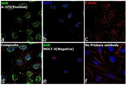
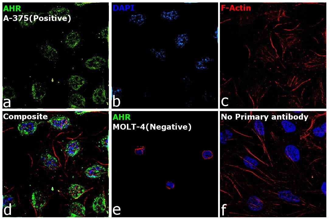
- Immunofluorescence analysis of AHR was performed using 70% confluent log phase A-375 cells. The cells were fixed with 4% Paraformaldehyde for 10 Minutes, permeabilized with 0.1% Triton™ X-100 for 10 Minutes, and blocked with 2% BSA for 10 Minutes at room temperature. The cells were labeled with AHR Monoclonal Antibody (FF3399), eBioscience (Product # 14-9854-82) at 20 µg/mL in 0.1% BSA, incubated at 4 degree Celsius overnight and then labeled with Goat anti-Mouse IgG (H+L), Superclonal™ Recombinant Secondary Antibody, Alexa Fluor 488 (Product # A28175), (1:2,000 dilution) for 45 minutes at room temperature (Panel a: Green). Nuclei (Panel b: Blue) were stained with SlowFade® Gold Antifade Mountant with DAPI (Product # S36938). F-actin (Panel c: Red) was stained with Rhodamine Phalloidin (Product # R415, 1:300). Panel d represents the merged image showing Cytoplasmic and nuclear localization. Panel e represents MOLT-4 cells having no expression of AHR. Panel f represents control cells with no primary antibody to assess background. The images were captured at 60X magnification.

- Submitted by
- Invitrogen Antibodies (provider)
- Main image
- Experimental details
- Immunofluorescence analysis of AHR was performed using 70% confluent log phase A-375 cells. The cells were fixed with 4% Paraformaldehyde for 10 Minutes, permeabilized with 0.1% Triton™ X-100 for 10 Minutes, and blocked with 2% BSA for 10 Minutes at room temperature. The cells were labeled with AHR Monoclonal Antibody (FF3399), eBioscience (Product # 14-9854-82) at 20 µg/mL in 0.1% BSA, incubated at 4 degree Celsius overnight and then labeled with Goat anti-Mouse IgG (H+L), Superclonal™ Recombinant Secondary Antibody, Alexa Fluor 488 (Product # A28175), (1:2,000 dilution) for 45 minutes at room temperature (Panel a: Green). Nuclei (Panel b: Blue) were stained with SlowFade® Gold Antifade Mountant with DAPI (Product # S36938). F-actin (Panel c: Red) was stained with Rhodamine Phalloidin (Product # R415, 1:300). Panel d represents the merged image showing Cytoplasmic and nuclear localization. Panel e represents MOLT-4 cells having no expression of AHR. Panel f represents control cells with no primary antibody to assess background. The images were captured at 60X magnification.
Supportive validation
- Submitted by
- Invitrogen Antibodies (provider)
- Main image
- Experimental details
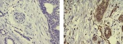
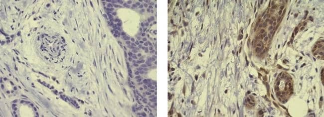
- Immunohistochemistry of formalin-fixed paraffin embedded human infiltrating ductal carcinoma using 20 µg/mL Mouse IgG2b K Isotype Control Purified (left) or 20 µg/mL Anti-Human AHR Purified (right) followed by Anti-Mouse IgG Biotin, Streptavidin HRP, and DAB visualization.Nuclei are counterstained with hematoxylin.
Supportive validation
- Submitted by
- Invitrogen Antibodies (provider)
- Main image

- Experimental details
- NULL
- Submitted by
- Invitrogen Antibodies (provider)
- Main image
- Experimental details
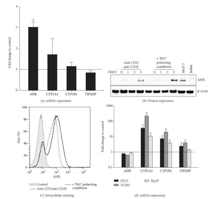
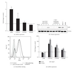
- Figure 5 The AHR is induced in CD4 + T cells and activated by FICZ, TCDD, and B[a]P. Human CD4 + T cells were cultured with anti-CD3/anti-CD28 in the presence or absence of Th17 polarizing conditions for 5 days. (a) The mRNA expression of the indicated genes was measured by real-time PCR. Data were normalized to the mean of the reference genes GUSB, PPIA, PGK1, and the mRNA expression at day 0 using the DeltaDeltaCt-method. Fold changes in gene expression of CD4 + T cells with Th17 polarizing conditions compared to control (black line) are represented as mean and SEM. *represents significance, P < 0.05, Mann-Whitney U test, and n = 7. (b) The AHR protein expression (94 kDa) at days 1, 3, and 5 of culture was measured by Western Blot analysis. The protein expression of unstimulated CD4 + T cells was used as control. Beta-Actin (42 kDa) antibody was used as loading control. Cell lysates from MCF-7 cell line and Jurkat T cell line were used as AHR positive and negative control, respectively. One representative of 3 independent experiments is shown. (c) After restimulation with PMA, Ionomycin, and Monensin for 5 hours and fixation/permeabilization, the cells were stained for CD4 and AHR and analyzed by flow cytometry. Unstained cells were used as control n = 2. (d) The AHR ligands FICZ (100 nM), TCDD (5 nM), and B[a]P (100 nM) were added to CD4 + T cells cultured with Th17 polarizing cytokines. CD4 + T cells treated with anti-CD3/anti-CD28, Th17 polarizing conditions, and DMSO (0