 Explore
 Validate
 Learn
Learn Western blot
 ELISA
ELISA Immunoprecipitation
Antibody data
- Antibody Data
- Antigen structure
- References [0]
- Comments [0]
- Validations
- Western blot [1]
- Immunoprecipitation [2]
Submit
Validation data
Reference
Comment
Report error
- Product number
- LS-C817750 - Provider product page

- Provider
- LSBio
- Product name
- ACTR3 Antibody (Preservative Free) LS-C817750
- Antibody type
- Polyclonal
- Description
- Protein A affinity chromatography
- Reactivity
- Human
- Host
- Rabbit
- Isotype
- IgG
- Storage
- Store at 2°C to 8°C for up to 1 month. Aliquot and store at -20°C to -80°C for up to 1 year. Avoid freeze/thaw cycles.
No comments: Submit comment
Enhanced validation
- Submitted by
- LSBio (provider)
- Enhanced method
- Genetic validation
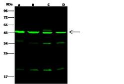
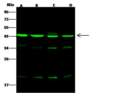
- Main image
- Experimental details
- Anti-ACTR3 rabbit polyclonal antibody at 1:500 dilution. Lane A: HeLa Whole Cell Lysate. Lane B: Jurkat Whole Cell Lysate. Lane C: A549 Whole Cell Lysate. Lane D: K562 Whole Cell Lysate. Lysates/proteins at 30 ug per lane. Secondary: Goat Anti-Rabbit IgG H&L (Dylight 800) at 1/10000 dilution. Developed using the Odyssey technique. Performed under reducing conditions. Predicted band size: 47 kDa. Observed band size: 45 kDa. (We are unsure as to the identity of these extra bands.)
Supportive validation
- Submitted by
- LSBio (provider)
- Enhanced method
- Genetic validation
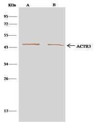
- Main image
- Experimental details
- ACTR3 was immunoprecipitated using: Lane A: 0.5 mg K562 Whole Cell Lysate. Lane B: 0.5 mg Hela Whole Cell Lysate. 4 uL anti-ACTR3 rabbit polyclonal antibody and 15 ul of 50% Protein G agarose. Primary antibody: Anti-ACTR3 rabbit polyclonal antibody, at 1:100 dilution. Secondary antibody: Clean-Blot IP Detection Reagent (HRP) at 1:500 dilution. Developed using the DAB staining technique. Performed under reducing conditions. Predicted band size: 47 kDa. Observed band size: 47 kDa.

- Submitted by
- LSBio (provider)
- Main image

- Experimental details
- ACTR3 was immunoprecipitated using: Lane A: 0.5 mg K562 Whole Cell Lysate. Lane B: 0.5 mg Hela Whole Cell Lysate. 4 uL anti-ACTR3 rabbit polyclonal antibody and 15 ul of 50% Protein G agarose. Primary antibody: Anti-ACTR3 rabbit polyclonal antibody, at 1:100 dilution. Secondary antibody: Clean-Blot IP Detection Reagent (HRP) at 1:500 dilution. Developed using the DAB staining technique. Performed under reducing conditions. Predicted band size: 47 kDa. Observed band size: 47 kDa.
