Antibody data
- Antibody Data
- Antigen structure
- References [1]
- Comments [0]
- Validations
- Western blot [4]
- Immunohistochemistry [1]
- Other assay [1]
Submit
Validation data
Reference
Comment
Report error
- Product number
- PA5-30354 - Provider product page
- Provider
- Invitrogen Antibodies
- Product name
- Arp3 Polyclonal Antibody
- Antibody type
- Polyclonal
- Antigen
- Recombinant protein fragment
- Description
- Recommended positive controls: Jurkat. Predicted reactivity: Mouse (100%), Rat (100%), Zebrafish (98%), Xenopus laevis (99%), Pig (99%), Chicken (99%), Chimpanzee (100%), Bovine (100%). Store product as a concentrated solution. Centrifuge briefly prior to opening the vial.
- Reactivity
- Human, Mouse, Rat
- Host
- Rabbit
- Isotype
- IgG
- Vial size
- 100 µL
- Concentration
- 1 mg/mL
- Storage
- Store at 4°C short term. For long term storage, store at -20°C, avoiding freeze/thaw cycles.
Submitted references Comparative 2D-DIGE proteomic analysis of bovine mammary epithelial cells during lactation reveals protein signatures for lactation persistency and milk yield.
Janjanam J, Singh S, Jena MK, Varshney N, Kola S, Kumar S, Kaushik JK, Grover S, Dang AK, Mukesh M, Prakash BS, Mohanty AK
PloS one 2014;9(8):e102515
No comments: Submit comment
Supportive validation
- Submitted by
- Invitrogen Antibodies (provider)
- Main image
- Experimental details
- Western Blot using Arp3 Polyclonal Antibody (Product # PA5-30354). Sample (30 µg of whole cell lysate). Lane A: Jurkat. 10% SDS PAGE. Arp3 Polyclonal Antibody (Product # PA5-30354) diluted at 1:1,000.
- Submitted by
- Invitrogen Antibodies (provider)
- Main image
- Experimental details
- Western Blot using Arp3 Polyclonal Antibody (Product # PA5-30354). Mouse tissue extract (50 µg) was separated by 10% SDS-PAGE, and the membrane was blotted with Arp3 Polyclonal Antibody (Product # PA5-30354) diluted at 1:1,000. The HRP-conjugated anti-rabbit IgG antibody was used to detect the primary antibody.
- Submitted by
- Invitrogen Antibodies (provider)
- Main image
- Experimental details
- Knockdown of Arp3 was achieved by transfecting MCF-7 cells with Arp3 specific siRNAs (Silencer® select Product # s19640, s19641). Western blot analysis (Fig. a) was performed using whole cell extracts from the MCF-7 knockdown cells (lane 3), non-specific scrambled siRNA transfected cells (lane 2) and untransfected cells (lane 1). The blot was probed with Arp3 Rabbit Polyclonal Antibody (Product # PA5-30354, 1:5000 dilution) and Goat anti-Rabbit IgG (H+L), Superclonal™ Recombinant Secondary Antibody, HRP (Product # A27036, 1:4000 dilution). Densitometric analysis of this western blot is shown in histogram (Fig. b). Decrease in signal upon siRNA mediated knock down confirms that antibody is specific to Arp3.
- Submitted by
- Invitrogen Antibodies (provider)
- Main image
- Experimental details
- Western blot was performed using Anti-Arp3 Polyclonal Antibody (Product # PA5-30354) and a 47 kDa band was observed across the panel tested except Mouse and Rat Skeletal Muscle which are reported to be negative. Whole cell extracts (30 µg lysate) of MDA-MB-231 (Lane 1), MCF-7 (Lane 2), PC-3 (Lane 3), U-87 MG (Lane 4), HeLa (Lane 5), U-2 OS (Lane 6), HEL 92.1.7 (Lane 7), tissue extracts of Mouse Thymus (Lane 8), Mouse Skeletal Muscle (Lane 9), Rat Thymus (Lane 10) and Rat Skeletal Muscle (Lane 11) were electrophoresed using NuPAGE™ 10% Bis-Tris Protein Gel (Product # NP0302BOX). Resolved proteins were then transferred onto a nitrocellulose membrane (Product # IB23001) by iBlot® 2 Dry Blotting System (Product # IB21001). The blot was probed with the primary antibody (1:1000 dilution) and detected by chemiluminescence with Goat anti-Rabbit IgG (H+L), Superclonal™ Recombinant Secondary Antibody, HRP (Product # A27036, 1:4000 dilution) using the iBright FL 1000 (Product # A32752). Chemiluminescent detection was performed using Novex® ECL Chemiluminescent Substrate Reagent Kit (Product # WP20005).
Supportive validation
- Submitted by
- Invitrogen Antibodies (provider)
- Main image
- Experimental details
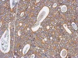
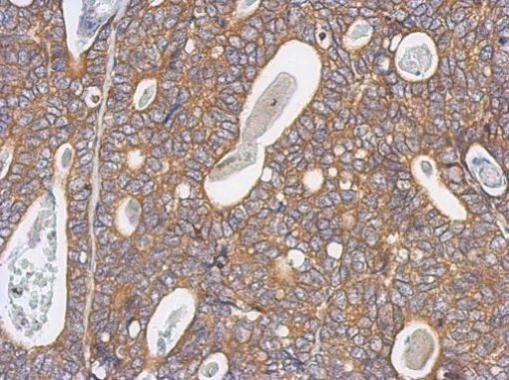
- Immunohistochemical analysis of paraffin-embedded human gastric cancer, using Arp3 (Product # PA5-30354) antibody at 1:500 dilution. Antigen Retrieval: EDTA based buffer, pH 8.0, 15 min.
Supportive validation
- Submitted by
- Invitrogen Antibodies (provider)
- Main image
- Experimental details
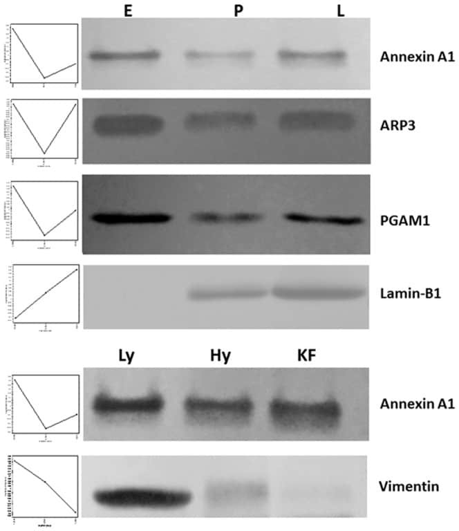
- Figure 11 Validation of 2D-DIGE data by western blot. Western blot analysis of annexin A1, ARP3, PGAM1 and Lamin-B1 proteins were differentially regulated during lactation stages and annexin A1 and vimentin were differentially regulated in high and low-milk yielding samples.