 Explore
 Validate
 Learn
Learn Western blot
 Immunoprecipitation
Antibody data
- Antibody Data
- Antigen structure
- References [0]
- Comments [0]
- Validations
- Western blot [3]
- Immunocytochemistry [1]
- Immunohistochemistry [1]
Submit
Validation data
Reference
Comment
Report error
- Product number
- PA5-47268 - Provider product page

- Provider
- Invitrogen Antibodies
- Product name
- Carbonic Anhydrase IX Polyclonal Antibody
- Antibody type
- Polyclonal
- Antigen
- Recombinant full-length protein
- Description
- In direct ELISAs, approximately 10% cross-reactivity with recombinant mouse CA9 is observed and less than 1% cross-reactivity with recombinant human CA1, 3, 4, 8, 10, 12, and 14 is observed. Reconstitute at 0.2 mg/mL in sterile PBS.
- Reactivity
- Human
- Host
- Goat
- Isotype
- IgG
- Vial size
- 100 µg
- Concentration
- 0.2 mg/mL
- Storage
- -20° C, Avoid Freeze/Thaw Cycles
No comments: Submit comment
Supportive validation
- Submitted by
- Invitrogen Antibodies (provider)
- Main image
- Experimental details
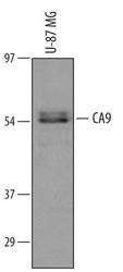
- Western blot analysis from lysates of U-87 MG human glioblastoma/astrocytoma cell line. PVDF membrane was probed with 1 µg/mL of Goat Anti-human Carbonic Anhydrase IX/CA9 Antigen Affinity-purified Polyclonal Antibody (Product # PA5-47268) followed by HRP-conjugated Anti-Goat IgG Secondary Antibody. A specific band was detected for Carbonic Anhydrase IX/CA9 at approximately 58 kDa (as indicated). This experiment was conducted under reducing conditions.
- Submitted by
- Invitrogen Antibodies (provider)
- Main image

- Experimental details
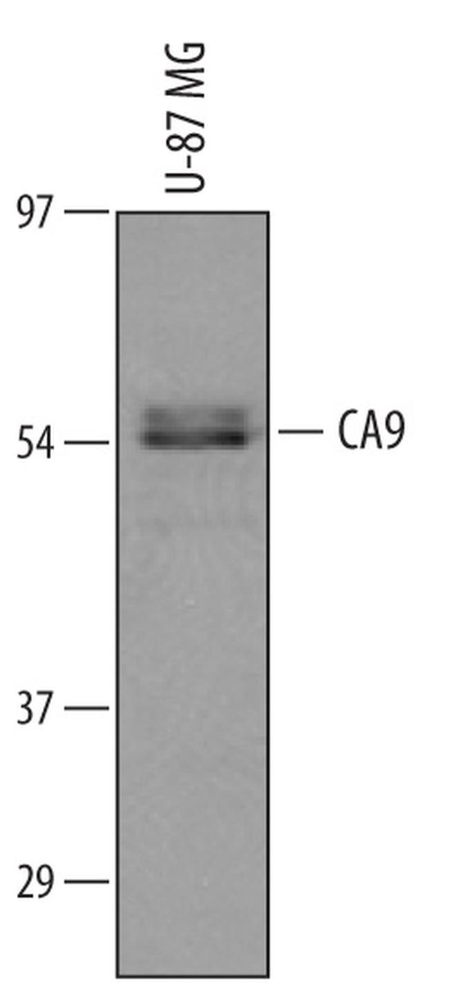
- Western blot analysis of Carbonic Anhydrase IX in U‚87 MG human glioblastoma/astrocytoma cell line. Samples were incubated in Carbonic Anhydrase IX polyclonal antibody (Product # PA5-47268) using a dilution of 1 µg/mL followed by a HRP-conjugated Anti-Goat IgG secondary antibody. A specific band was detected for Carbonic Anhydrase IX/CA9 at approximately 58 kDa (as indicated). This experiment was conducted under reducing conditions.

- Submitted by
- Invitrogen Antibodies (provider)
- Main image

- Experimental details
- Western blot was performed using Anti-Carbonic Anhydrase IX Polyclonal Antibody (Product # PA5-47268) and ~ 50-55 kDa bands corresponding to CA9 was observed across cell lines tested and increased upon Deferoxamine and Cobalt Chloride treatment in HeLa. Whole cell extracts (30 µg lysate) of U-87 MG (Lane 1), HeLa (Lane 2), HeLa treated with Deferoxamine (100uM for 24 Hours) (Lane 3) and HeLa treated with Cobalt Chloride (200uM for 24 Hours) (Lane 4) were electrophoresed using NuPAGE® 4-12 % Bis-Tris gel (Product # NP0322BOX). Resolved proteins were then transferred onto a nitrocellulose membrane (Product # IB23001) by iBlot® 2 Dry Blotting System (Product # IB21001). The blot was probed with the primary antibody (1ug/ml) and detected by chemiluminescence with Rabbit anti-Goat IgG (H+L), Superclonal™ Recombinant Secondary Antibody, HRP (Product # A27014, 1:4000 dilution) using the iBright FL 1000 (Product # A32752). Chemiluminescent detection was performed using Novex® ECL Chemiluminescent Substrate Reagent Kit (Product # WP20005).

Supportive validation
- Submitted by
- Invitrogen Antibodies (provider)
- Main image

- Experimental details
- Immunocytochemistry analysis of Carbonic Anhydrase IX in immersion fixed A431 human epithelial carcinoma cell line. Samples were incubated in Carbonic Anhydrase IX polyclonal antibody (Product # PA5-47268) using a dilution of 3 µg/mL for 3 hours at room temperature followed by NorthernLights™ 557-conjugated Anti-Goat IgG Secondary Antibody (red) and counterstained with DAPI (blue). Specific staining was localized to cytoplasm.

Supportive validation
- Submitted by
- Invitrogen Antibodies (provider)
- Main image

- Experimental details
- Immunohistochemical analysis of Carbonic Anhydrase IX in immersion fixed paraffin-embedded sections of human colon cancer tissue. Samples were incubated in Carbonic Anhydrase IX polyclonal antibody (Product # PA5-47268) using a dilution of 15 µg/mL overnight at 4 °C. Tissue was stained using the Anti-Goat HRP-DAB Cell & Tissue Staining Kit (brown) and counterstained with hematoxylin (blue). Specific labeling was localized to the plasma membrane of epithelial cells.
