 Explore
 Validate
 Learn
Learn Western blot
 Immunoprecipitation
Antibody data
- Antibody Data
- Antigen structure
- References [3]
- Comments [0]
- Validations
- Western blot [1]
- Immunocytochemistry [1]
- Immunohistochemistry [1]
Submit
Validation data
Reference
Comment
Report error
- Product number
- AF2188 - Provider product page

- Provider
- R&D Systems
- Product name
- Human Carbonic Anhydrase IX/CA9 Antibody
- Antibody type
- Polyclonal
- Description
- Antigen Affinity-purified. Detects human Carbonic Anhydrase IX (CA9) in direct ELISAs and Western blots. In direct ELISAs, approximately 10% cross-reactivity with recombinant mouse CA9 is observed and less than 1% cross-reactivity with recombinant human CA1, 3, 4, 8, 10, 12, and 14 is observed.
- Reactivity
- Human
- Host
- Goat
- Conjugate
- Unconjugated
- Antigen sequence
Q16790- Isotype
- IgG
- Vial size
- 100 ug
- Concentration
- LYOPH
- Storage
- Use a manual defrost freezer and avoid repeated freeze-thaw cycles. 12 months from date of receipt, -20 to -70 °C as supplied. 1 month, 2 to 8 °C under sterile conditions after reconstitution. 6 months, -20 to -70 °C under sterile conditions after reconstitution.
Submitted references Activation of the hypoxia pathway in breast cancer tissue and patient survival are inversely associated with tumor ascorbate levels.
Compensation of Signal Spillover in Suspension and Imaging Mass Cytometry.
The interactome of metabolic enzyme carbonic anhydrase IX reveals novel roles in tumor cell migration and invadopodia/MMP14-mediated invasion.
Campbell EJ, Dachs GU, Morrin HR, Davey VC, Robinson BA, Vissers MCM
BMC cancer 2019 Apr 3;19(1):307
BMC cancer 2019 Apr 3;19(1):307
Compensation of Signal Spillover in Suspension and Imaging Mass Cytometry.
Chevrier S, Crowell HL, Zanotelli VRT, Engler S, Robinson MD, Bodenmiller B
Cell systems 2018 May 23;6(5):612-620.e5
Cell systems 2018 May 23;6(5):612-620.e5
The interactome of metabolic enzyme carbonic anhydrase IX reveals novel roles in tumor cell migration and invadopodia/MMP14-mediated invasion.
Swayampakula M, McDonald PC, Vallejo M, Coyaud E, Chafe SC, Westerback A, Venkateswaran G, Shankar J, Gao G, Laurent EMN, Lou Y, Bennewith KL, Supuran CT, Nabi IR, Raught B, Dedhar S
Oncogene 2017 Nov 9;36(45):6244-6261
Oncogene 2017 Nov 9;36(45):6244-6261
No comments: Submit comment
Supportive validation
- Submitted by
- R&D Systems (provider)
- Main image
- Experimental details
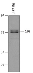
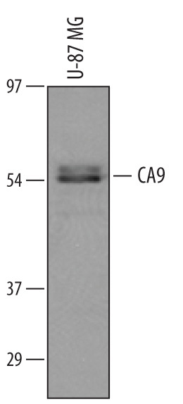
- Detection of Human Carbonic Anhydrase IX/CA9 by Western Blot. Western blot shows lysates of U-87 MG human glioblastoma/ astrocytoma cell line. PVDF membrane was probed with 1 µg/mL of Goat Anti-Human Carbonic Anhydrase IX/CA9 Antigen Affinity-purified Polyclonal Antibody (Catalog # AF2188) followed by HRP-conjugated Anti-Goat IgG Secondary Antibody (Catalog # HAF109). A specific band was detected for Carbonic Anhydrase IX/CA9 at approximately 58 kDa (as indicated). This experiment was conducted under reducing conditions and using Immunoblot Buffer Group 8.
Supportive validation
- Submitted by
- R&D Systems (provider)
- Main image
- Experimental details
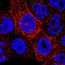
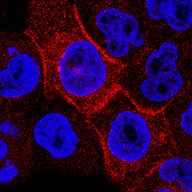
- Carbonic Anhydrase IX/CA9 in A431 Human Cell Line. Carbonic Anhydrase IX/CA9 was detected in immersion fixed A431 human epithelial carcinoma cell line using Goat Anti-Human Carbonic Anhydrase IX/CA9 Antigen Affinity-purified Polyclonal Antibody (Catalog # AF2188) at 3 µg/mL for 3 hours at room temperature. Cells were stained using the NorthernLights™ 557-conjugated Anti-Goat IgG Secondary Antibody (red; Catalog # NL001) and counterstained with DAPI (blue). Specific staining was localized to cytoplasm. View our protocol for Fluorescent ICC Staining of Cells on Coverslips.
Supportive validation
- Submitted by
- R&D Systems (provider)
- Main image
- Experimental details
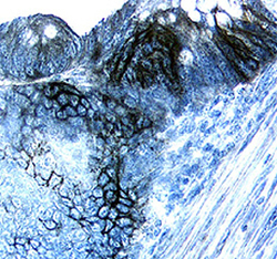
- Carbonic Anhydrase IX/CA9 in Human Colon Cancer Tissue. Carbonic Anhydrase IX/CA9 was detected in immersion fixed paraffin-embedded sections of human colon cancer tissue using Goat Anti-Human Carbonic Anhydrase IX/CA9 Antigen Affinity-purified Polyclonal Antibody (Catalog # AF2188) at 15 µg/mL overnight at 4 °C. Tissue was stained using the Anti-Goat HRP-DAB Cell & Tissue Staining Kit (brown; Catalog # CTS008) and counterstained with hematoxylin (blue). Specific labeling was localized to the plasma membrane of epithelial cells. View our protocol for Chromogenic IHC Staining of Paraffin-embedded Tissue Sections.
