 Explore
 Validate
 Learn
Learn Western blot
Antibody data
- Antibody Data
- Antigen structure
- References [0]
- Comments [0]
- Validations
- Western blot [2]
- Immunocytochemistry [1]
- Immunohistochemistry [1]
Submit
Validation data
Reference
Comment
Report error
- Product number
- PA1-16634 - Provider product page

- Provider
- Invitrogen Antibodies
- Product name
- CTGF Polyclonal Antibody
- Antibody type
- Polyclonal
- Antigen
- Synthetic peptide
- Description
- This antibody is predicted to react with porcine, bovine, chicken and sheep based on 100% sequence homology.
- Concentration
- 1.0 mg/mL
No comments: Submit comment
Supportive validation
- Submitted by
- Invitrogen Antibodies (provider)
- Main image

- Experimental details
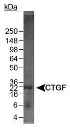
- Detection of CTGF in mouse kidney lysate using Product # PA1-16634. 5 sec. ECL exposure.
- Submitted by
- Invitrogen Antibodies (provider)
- Main image

- Experimental details
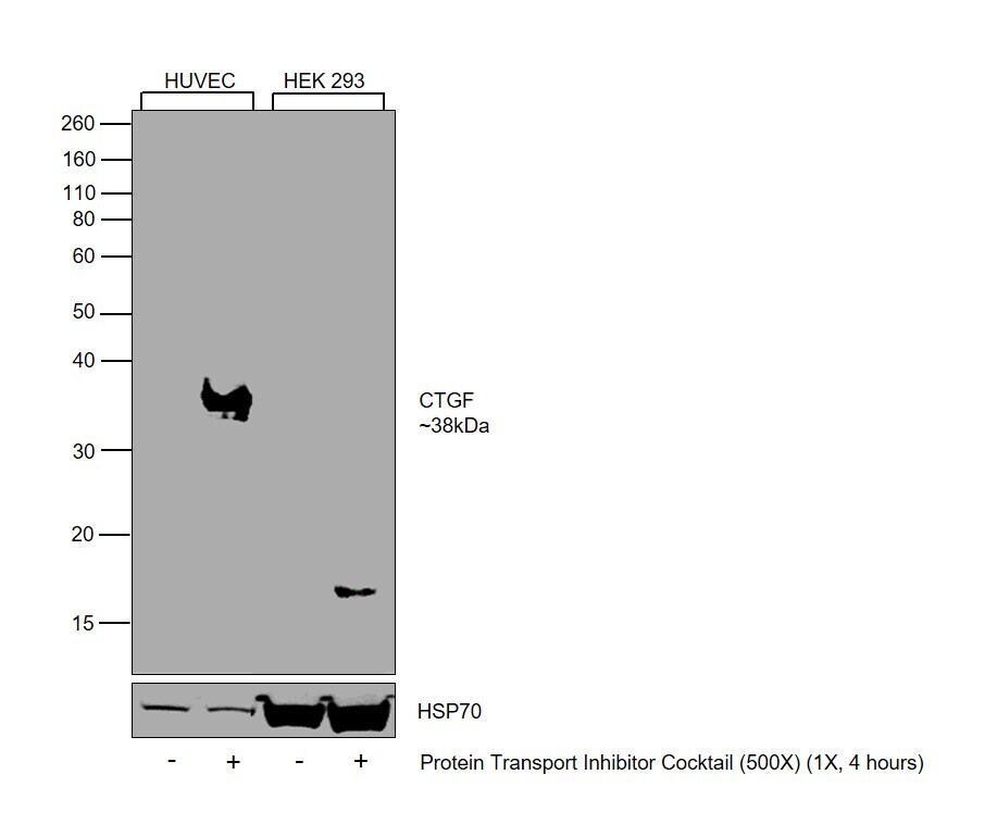
- Western Blot was performed using Anti-CTGF Polyclonal Antibody (Product # PA1-16634) and a 38 kDa band corresponding to Connective tissue growth factor was observed across in HUVEC upon PTI treatment, but not in HEK 293 upon PTI treatment. Whole cell extracts (30 µg lysate) of HUVEC (Lane 1), HUVEC (Protein Transport Inhibitor Cocktail (500X), 1X, 4 hours) (Lane 2), HEK-293 (Lane 3), HEK-293 (Protein Transport Inhibitor Cocktail (500X), 1X, 4 hours) (Lane 4) were electrophoresed using NuPAGE™ 10% Bis-Tris Protein Gel (Product # NP0302BOX). Resolved proteins were then transferred onto a Nitrocellulose membrane (Product # IB23001) by iBlot® 2 Dry Blotting System (Product # IB21001). The Blot was probed with the primary antibody (1:1000) and detected by chemiluminescence with Goat anti-Rabbit IgG (H+L) Superclonal™ Recombinant Secondary Antibody, HRP (Product # A27036, 1:4000) using the iBright FL 1000 (Product # A32752). Chemiluminescent detection was performed using SuperSignal™ West Dura Extended Duration Substrate (Product # 34076).
Supportive validation
- Submitted by
- Invitrogen Antibodies (provider)
- Main image
- Experimental details
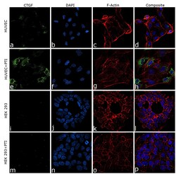
- Immunofluorescence analysis of CTGF was performed using 70% confluent log phase HUVEC cells. The cells were fixed with 4% paraformaldehyde for 10 minutes, permeabilized with 0.1% Triton™ X-100 for 15 minutes, and blocked with 2% BSA for 45 minutes at room temperature. The cells were labeled with CTGF Polyclonal Antibody (Product # PA1-16634) at 1:100 in 0.1% BSA, incubated at 4 degree celsius overnight and then labeled with Donkey anti-Rabbit IgG (H+L) Highly Cross-Adsorbed Secondary Antibody, Alexa Fluor Plus 488 (Product # A32790), (1:2000), for 45 minutes at room temperature (Panel a: Green). Nuclei (Panel b: Blue) were stained with ProLong™ Diamond Antifade Mountant with DAPI (Product # P36962). F-actin (Panel c: Red) was stained with Rhodamine Phalloidin (Product # R415, 1:300). Panel d represents the merged image showing cytoplasmic localization. Panel (a-d) shows representative HUVEC control cell, whereas Panel (e-h) represent HUVEC with PTI treatment. Similarly, panel (i-l) represent HEK 293 control cells and Panel (m-p) represent HEK 293 with PTI treatment. The images were captured at 60X magnification.

Supportive validation
- Submitted by
- Invitrogen Antibodies (provider)
- Main image

- Experimental details
- Immunohistochemical analysis of CTGF in formalin-fixed paraffin-embedded tissue section of human lymph node. Samples were incubated in CTGF polyclonal antibody (Product # PA1-16634) using a dilution of 1:200 followed by HRP labeled anti-rabbit secondary antibody and DAB reagent. Nuclei of cells were counter-stained with hematoxylin.
