Explore
 Validate
 Learn
Learn Western blot
 Immunocytochemistry
Antibody data
- Antibody Data
- Antigen structure
- References [3]
- Comments [0]
- Validations
- Immunocytochemistry [2]
- Immunohistochemistry [1]
- Other assay [1]
Submit
Validation data
Reference
Comment
Report error
- Product number
- PA5-32193 - Provider product page

- Provider
- Invitrogen Antibodies
- Product name
- CTGF Polyclonal Antibody
- Antibody type
- Polyclonal
- Antigen
- Recombinant full-length protein
- Description
- Recommended positive controls: A549, H1299, HCT116, HUVEC, Mouse heart, Rat bladder, CTGF-transfected 293T, HepG2, HepG2 conditioned medium. Predicted reactivity: Mouse (93%), Rat (94%), Xenopus laevis (85%), Pig (95%), Chicken (94%), Sheep (96%), Chimpanzee (99%), Bovine (93%). Store product as a concentrated solution. Centrifuge briefly prior to opening the vial.
- Reactivity
- Human, Mouse, Rat, Bovine
- Host
- Rabbit
- Isotype
- IgG
- Vial size
- 100 μL
- Concentration
- 0.42 mg/mL
- Storage
- Store at 4°C short term. For long term storage, store at -20°C, avoiding freeze/thaw cycles.
Submitted references Role of the Hippo signaling pathway in safflower yellow pigment treatment of paraquat-induced pulmonary fibrosis.
The mevalonate coordinates energy input and cell proliferation.
Synergistic Effect of Simvastatin Plus Radiation in Gastric Cancer and Colorectal Cancer: Implications of BIRC5 and Connective Tissue Growth Factor.
Li H, Kan B, Song L, Liu Y, Jian X
The Journal of international medical research 2020 Sep;48(9):300060520905425
The Journal of international medical research 2020 Sep;48(9):300060520905425
The mevalonate coordinates energy input and cell proliferation.
Gong L, Xiao Y, Xia F, Wu P, Zhao T, Xie S, Wang R, Wen Q, Zhou W, Xu H, Zhu L, Zheng Z, Yang T, Chen Z, Duan Q
Cell death & disease 2019 Apr 11;10(4):327
Cell death & disease 2019 Apr 11;10(4):327
Synergistic Effect of Simvastatin Plus Radiation in Gastric Cancer and Colorectal Cancer: Implications of BIRC5 and Connective Tissue Growth Factor.
Lim T, Lee I, Kim J, Kang WK
International journal of radiation oncology, biology, physics 2015 Oct 1;93(2):316-25
International journal of radiation oncology, biology, physics 2015 Oct 1;93(2):316-25
No comments: Submit comment
Supportive validation
- Submitted by
- Invitrogen Antibodies (provider)
- Main image
- Experimental details
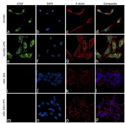
- Immunofluorescence analysis of CTGF was performed using 70% confluent log phase HUVEC cells. The cells were fixed with 4% paraformaldehyde for 10 minutes, permeabilized with 0.1% Triton™ X-100 for 10 minutes, and blocked with 2% BSA for 10 minutes at room temperature. The cells were labeled with CTGF Polyclonal Antibody (Product # PA5-32193) at 5 µg/mL in 0.1% BSA, incubated at 4 degree celsius overnight and then labeled with Donkey anti-Rabbit IgG (H+L) Highly Cross-Adsorbed Secondary Antibody, Alexa Fluor Plus 488 (Product # A32790), (1:2000 dilution), for 45 minutes at room temperature (Panel a: Green). Nuclei (Panel b:Blue) were stained with ProLong™ Diamond Antifade Mountant with DAPI (Product # P36962). F-actin (Panel c: Red) was stained with Rhodamine Phalloidin (Product # R415, 1:300). Panel d represents the merged image showing Cytoplasmic localization. Panel (a-d) shows representative HUVEC control cell, whereas Panel (e-h) represent HUVEC cells treated with Protein Transport Inhibitor Cocktail (500X), 1X, 4 hours. Similarly, panel (i-l) represent HEK 293 control cells and Panel (m-p) represent HEK 293 treated with Protein Transport Inhibitor Cocktail (500X), 1X, 4 hours. The images were captured at 60X magnification.

- Submitted by
- Invitrogen Antibodies (provider)
- Main image

- Experimental details
- Immunofluorescence analysis of CTGF was performed using 70% confluent log phase HUVEC cells. The cells were fixed with 4% paraformaldehyde for 10 minutes, permeabilized with 0.1% Triton™ X-100 for 10 minutes, and blocked with 2% BSA for 10 minutes at room temperature. The cells were labeled with CTGF Polyclonal Antibody (Product # PA5-32193) at 5 µg/mL in 0.1% BSA, incubated at 4 degree celsius overnight and then labeled with Donkey anti-Rabbit IgG (H+L) Highly Cross-Adsorbed Secondary Antibody, Alexa Fluor Plus 488 (Product # A32790), (1:2000 dilution), for 45 minutes at room temperature (Panel a: Green). Nuclei (Panel b:Blue) were stained with ProLong™ Diamond Antifade Mountant with DAPI (Product # P36962). F-actin (Panel c: Red) was stained with Rhodamine Phalloidin (Product # R415, 1:300). Panel d represents the merged image showing Cytoplasmic localization. Panel (a-d) shows representative HUVEC control cell, whereas Panel (e-h) represent HUVEC cells treated with Protein Transport Inhibitor Cocktail (500X), 1X, 4 hours. Similarly, panel (i-l) represent HEK 293 control cells and Panel (m-p) represent HEK 293 treated with Protein Transport Inhibitor Cocktail (500X), 1X, 4 hours. The images were captured at 60X magnification.

Supportive validation
- Submitted by
- Invitrogen Antibodies (provider)
- Main image
- Experimental details
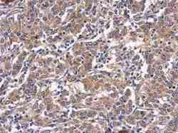
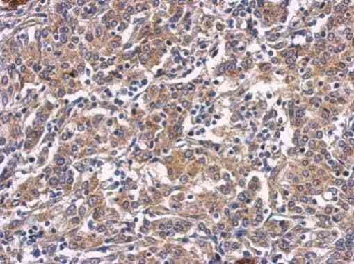
- Immunohistochemical analysis of paraffin-embedded human gastric cancer, using CTGF (Product # PA5-32193) antibody at 1:500 dilution. Antigen Retrieval: EDTA based buffer, pH 8.0, 15 min.
Supportive validation
- Submitted by
- Invitrogen Antibodies (provider)
- Main image
- Experimental details
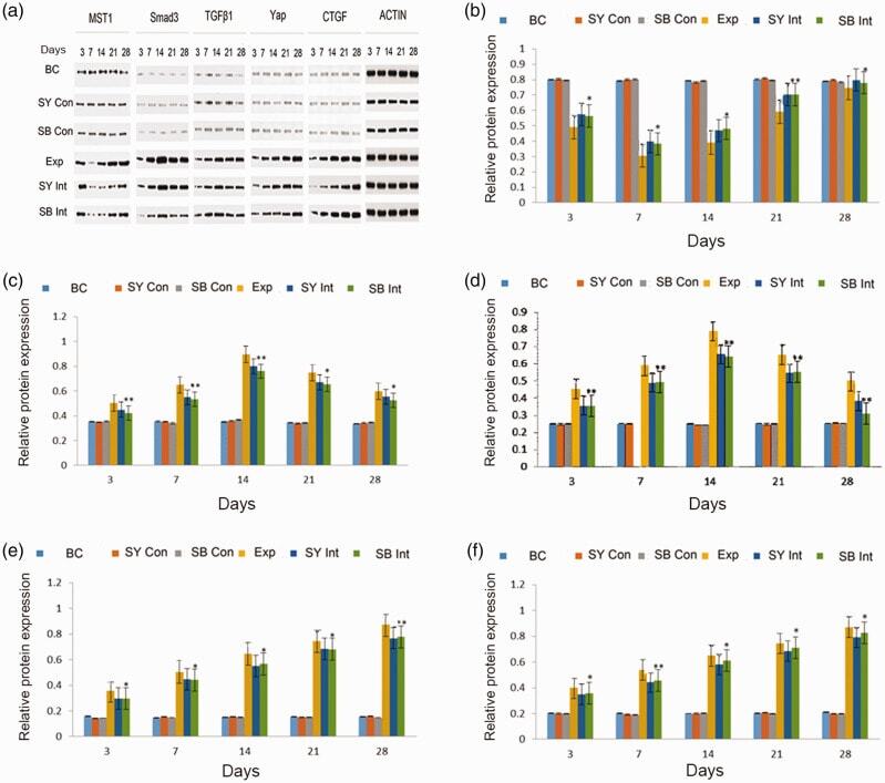
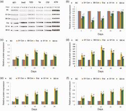
- Figure 4. Immunoblotting analysis of MST, TGF-beta1, Smad3, Yap and CTGF among groups over time. Actin was used as an internal reference. a. Protein expression levels of MST, TGF-beta1, Smad3, Yap, CTGF and actin. b. Quantification of MST protein expression in lung tissue. c. Quantification of Smad3 protein expression in lung tissue. d. Quantification of TGF-beta1 protein expression in lung tissue. e. Quantification of Yap protein expression in lung tissue. f. Quantification of CTGF protein expression in lung tissue. Exp vs BC: aa (P < 0.01), a (P < 0.05); Exp vs SY Int: bb (P < 0.01), b (P < 0.05); Exp vs SB Int: **(P < 0.01), *(P < 0.05).Abbreviations: BC, blank control; SY Con, safflower yellow pigment control; SB Con, SB431542 control; Exp, exposure; SY Int, safflower yellow pigment intervention; SB Int, SB431542 intervention; MST, mammalian STE20-like; TGF-beta1, transforming growth factor-beta1; CTGF, connective tissue growth factor.