 Explore
 Validate
 Learn
Learn ELISA
ELISA Immunocytochemistry
Antibody data
- Antibody Data
- Antigen structure
- References [12]
- Comments [0]
- Validations
- Immunocytochemistry [1]
- Other assay [1]
Submit
Validation data
Reference
Comment
Report error
- Product number
- 33-8200 - Provider product page

- Provider
- Invitrogen Antibodies
- Product name
- Cytochrome C Monoclonal Antibody (6H2.B4)
- Antibody type
- Monoclonal
- Antigen
- Other
- Description
- This antibody reacts with native Cytochrome C, but does not recognize denatured cytochrome C. The antibody recognizes a region in the vicinity of amino acid 62 of rat Cytochrome C.
- Reactivity
- Human, Mouse, Rat, Drosophila, Rabbit
- Host
- Mouse
- Isotype
- IgG
- Antibody clone number
- 6H2.B4
- Vial size
- 100 µg
- Concentration
- 0.5 mg/mL
- Storage
- Maintain refrigerated at 2-8°C for up to 1 month. For long term storage store at -20°C
Submitted references Enhancing Proteotoxic Stress in Leiomyosarcoma Cells Triggers Mitochondrial Dysfunctions, Cell Death, and Antitumor Activity in vivo.
Degradation of altered mitochondria by autophagy is impaired in Lafora disease.
Intracellular glycolysis in brown adipose tissue is essential for optogenetically induced nonshivering thermogenesis in mice.
Actomyosin drives cancer cell nuclear dysmorphia and threatens genome stability.
Loss of outer retinal neurons and circuitry alterations in the DBA/2J mouse.
Parkinson's disease transgenic mitochondrial cybrids generate Lewy inclusion bodies.
Parkinson's disease transgenic mitochondrial cybrids generate Lewy inclusion bodies.
Afferent regulation of cytochrome-c and active caspase-9 in the avian cochlear nucleus.
Mitochondrial and nucleocytoplasmic targeting of O-linked GlcNAc transferase.
Mitochondrial and nucleocytoplasmic targeting of O-linked GlcNAc transferase.
The Toxoplasma gondii protein ROP2 mediates host organelle association with the parasitophorous vacuole membrane.
Elevated AKT activity protects the prostate cancer cell line LNCaP from TRAIL-induced apoptosis.
Iuliano L, Drioli S, Pignochino Y, Cafiero CM, Minisini M, D'Este F, Picco R, Dalla E, Giordano G, Grignani G, Di Giorgio E, Benedetti F, Felluga F, Brancolini C
Molecular cancer therapeutics 2021 Jun;20(6):1039-1051
Molecular cancer therapeutics 2021 Jun;20(6):1039-1051
Degradation of altered mitochondria by autophagy is impaired in Lafora disease.
Lahuerta M, Aguado C, Sánchez-Martín P, Sanz P, Knecht E
The FEBS journal 2018 Jun;285(11):2071-2090
The FEBS journal 2018 Jun;285(11):2071-2090
Intracellular glycolysis in brown adipose tissue is essential for optogenetically induced nonshivering thermogenesis in mice.
Jeong JH, Chang JS, Jo YH
Scientific reports 2018 Apr 27;8(1):6672
Scientific reports 2018 Apr 27;8(1):6672
Actomyosin drives cancer cell nuclear dysmorphia and threatens genome stability.
Takaki T, Montagner M, Serres MP, Le Berre M, Russell M, Collinson L, Szuhai K, Howell M, Boulton SJ, Sahai E, Petronczki M
Nature communications 2017 Jul 24;8:16013
Nature communications 2017 Jul 24;8:16013
Loss of outer retinal neurons and circuitry alterations in the DBA/2J mouse.
Fernández-Sánchez L, de Sevilla Müller LP, Brecha NC, Cuenca N
Investigative ophthalmology & visual science 2014 Aug 12;55(9):6059-72
Investigative ophthalmology & visual science 2014 Aug 12;55(9):6059-72
Parkinson's disease transgenic mitochondrial cybrids generate Lewy inclusion bodies.
Trimmer PA, Borland MK, Keeney PM, Bennett JP Jr, Parker WD Jr
Journal of neurochemistry 2004 Feb;88(4):800-12
Journal of neurochemistry 2004 Feb;88(4):800-12
Parkinson's disease transgenic mitochondrial cybrids generate Lewy inclusion bodies.
Trimmer PA, Borland MK, Keeney PM, Bennett JP Jr, Parker WD Jr
Journal of neurochemistry 2004 Feb;88(4):800-12
Journal of neurochemistry 2004 Feb;88(4):800-12
Afferent regulation of cytochrome-c and active caspase-9 in the avian cochlear nucleus.
Wilkinson BL, Elam JS, Fadool DA, Hyson RL
Neuroscience 2003;120(4):1071-9
Neuroscience 2003;120(4):1071-9
Mitochondrial and nucleocytoplasmic targeting of O-linked GlcNAc transferase.
Love DC, Kochan J, Cathey RL, Shin SH, Hanover JA
Journal of cell science 2003 Feb 15;116(Pt 4):647-54
Journal of cell science 2003 Feb 15;116(Pt 4):647-54
Mitochondrial and nucleocytoplasmic targeting of O-linked GlcNAc transferase.
Love DC, Kochan J, Cathey RL, Shin SH, Hanover JA
Journal of cell science 2003 Feb 15;116(Pt 4):647-54
Journal of cell science 2003 Feb 15;116(Pt 4):647-54
The Toxoplasma gondii protein ROP2 mediates host organelle association with the parasitophorous vacuole membrane.
Sinai AP, Joiner KA
The Journal of cell biology 2001 Jul 9;154(1):95-108
The Journal of cell biology 2001 Jul 9;154(1):95-108
Elevated AKT activity protects the prostate cancer cell line LNCaP from TRAIL-induced apoptosis.
Nesterov A, Lu X, Johnson M, Miller GJ, Ivashchenko Y, Kraft AS
The Journal of biological chemistry 2001 Apr 6;276(14):10767-74
The Journal of biological chemistry 2001 Apr 6;276(14):10767-74
No comments: Submit comment
Supportive validation
- Submitted by
- Invitrogen Antibodies (provider)
- Main image
- Experimental details
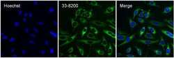
- Immunofluorescent analysis of Cytochrome C (green) in HeLa cells. The cells were fixed with 4% Paraformaldehyde in PBS for 15 minutes and blocked with 3% BSA in PBS for 30 minutes at room temperature. Cells were stained with a Cytochrome C mouse monoclonal antibody (Product # 33-8200) at a dilution of 5 µg/mL in blocking buffer for 1 hour at room temperature, and then incubated with a Goat anti-Mouse IgG (H+L) Superclonal™ Secondary Antibody, Alexa Fluor® 488 conjugate (Product # A28175) at a dilution of 1:1000 for 1 hour at room temperature. Nuclei (blue) were stained with Hoechst Dye (Product # 62249). Images were taken at 20X magnification on a Thermo Scientific ToxInsight HCS imaging instrument.

Supportive validation
- Submitted by
- Invitrogen Antibodies (provider)
- Main image

- Experimental details
- Figure 6 Blockade of MCT1 inhibits the effects of optogenetic stimulation of sympathetic efferent fibers of BAT. ( a ) Cropped images of western blotting showing expression of MCT1 in the mitochondrial fractions (n = 2 mice). Full-length blots are presented in Supplementary Figure 2. VDAC: voltage-dependent anion channel, Cyt. C: cytochrome C, Cadhe: Cadherin ( b ) Pooled data from 6 mice showing changes in Slc16a1 ( Mct1 ) mRNA expression with (filled square) and without (open circle) stimulation of sympathetic innervation of BAT (***p < 0.001, unpaired t -test). Data are shown as mean +- SEM. ( c ) Pooled data showing effects of blockade of the MCT1 on optogenetically induced increase in body temperature and glucose uptake (n = 5 mice). Data are shown as mean +- SEM. ( d ) Plot showing relative Slc16a1 (Mct1) mRNA expression in mice injected with control or Slc16a1 shRNA into BAT (n = 6 mice, ***p < 0.001, unpaired t -test). Data are shown as mean +- SEM. ( e ) Western blotting to show knockdown of the Mct1 gene in BAT injected with Mct1 shRNA. Cropped images of western blotting showing knockdown of MCT1 protein (upper panel). Plot showing relative expression of MCT1 protein (bottom panel, *p < 0.05, unpaired t -test). Data are shown as mean +- SEM. ( f ) Pooled data showing that mice injected with Slc16a1 (Mct1) shRNA into the BAT pad showed no response to optogenetic stimulation. Data are shown as mean +- SEM.
