Explore
 Validate
 Learn
Learn Western blot
 Immunocytochemistry
Antibody data
- Antibody Data
- Antigen structure
- References [12]
- Comments [0]
- Validations
- Immunocytochemistry [4]
- Flow cytometry [2]
- Other assay [5]
Submit
Validation data
Reference
Comment
Report error
- Product number
- 45-6100 - Provider product page

- Provider
- Invitrogen Antibodies
- Product name
- Cytochrome C Monoclonal Antibody (37BA11)
- Antibody type
- Monoclonal
- Antigen
- Other
- Description
- 45-6100 was used successfully in the western blot analysis of cytochrome C using human breast cancer cell line lysates. Positive control: Isolated mitochondria from Human, Bovine, Rat and Mouse heart; Human fibroblasts; HeLa cells. WB: Predicted molecular weight: 14 kDa. ICC/IF: Heat induced antigen retrieval in 0.1 M Tris-HCl, 5% urea, pH 9.5 for 5 min at 95°C improves signal.
- Reactivity
- Human, Mouse, Rat, Bovine
- Host
- Mouse
- Isotype
- IgG
- Antibody clone number
- 37BA11
- Vial size
- 100 μL
- Concentration
- 1 mg/mL
- Storage
- -20°C, Avoid Freeze/Thaw Cycles
Submitted references Mechanical Study of Jian-Gan-Xiao-Zhi Decoction on Nonalcoholic Fatty Liver Disease Based on Integrated Network Pharmacology and Untargeted Metabolomics.
MicroRNA-325-3p Facilitates Immune Escape of Mycobacterium tuberculosis through Targeting LNX1 via NEK6 Accumulation to Promote Anti-Apoptotic STAT3 Signaling.
16-Hydroxycleroda-3,13-dien-15,16-olide Induces Apoptosis in Human Bladder Cancer Cells through Cell Cycle Arrest, Mitochondria ROS Overproduction, and Inactivation of EGFR-Related Signalling Pathways.
DNA methylation and histone deacetylation regulating insulin sensitivity due to chronic cold exposure.
A ketogenic diet accelerates neurodegeneration in mice with induced mitochondrial DNA toxicity in the forebrain.
Evolutionarily conserved intercalated disc protein Tmem65 regulates cardiac conduction and connexin 43 function.
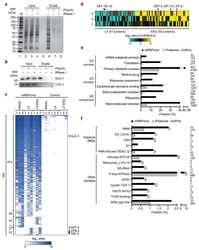
Conserved mRNA-binding proteomes in eukaryotic organisms.
Specific acetylation of p53 by HDAC inhibition prevents DNA damage-induced apoptosis in neurons.
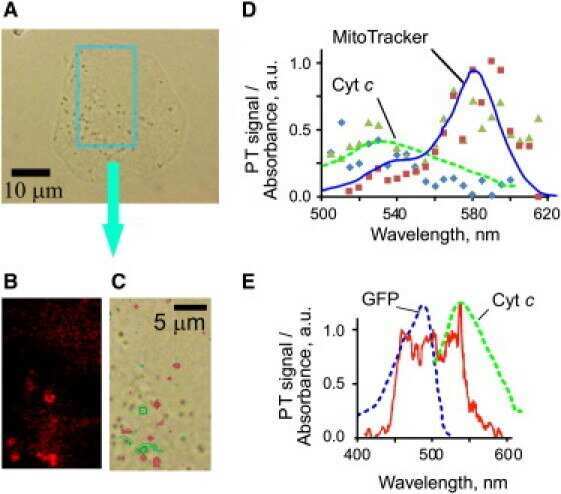
Photothermal confocal spectromicroscopy of multiple cellular chromophores and fluorophores.
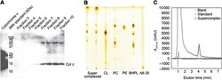
Arrangement of electron transport chain components in bovine mitochondrial supercomplex I1III2IV1.
Mitochondrial DNA toxicity compromises mitochondrial dynamics and induces hippocampal antioxidant defenses.
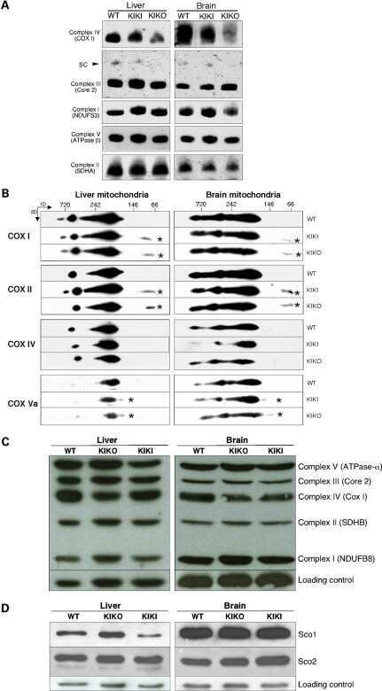
Analysis of mouse models of cytochrome c oxidase deficiency owing to mutations in Sco2.
Cao YJ, Li HZ, Zhao J, Sun YM, Jin XW, Lv SQ, Luo JY, Fang XX, Wen WB, Liao JB
Evidence-based complementary and alternative medicine : eCAM 2022;2022:2264394
Evidence-based complementary and alternative medicine : eCAM 2022;2022:2264394
MicroRNA-325-3p Facilitates Immune Escape of Mycobacterium tuberculosis through Targeting LNX1 via NEK6 Accumulation to Promote Anti-Apoptotic STAT3 Signaling.
Fu B, Xue W, Zhang H, Zhang R, Feldman K, Zhao Q, Zhang S, Shi L, Pavani KC, Nian W, Lin X, Wu H
mBio 2020 Jun 2;11(3)
mBio 2020 Jun 2;11(3)
16-Hydroxycleroda-3,13-dien-15,16-olide Induces Apoptosis in Human Bladder Cancer Cells through Cell Cycle Arrest, Mitochondria ROS Overproduction, and Inactivation of EGFR-Related Signalling Pathways.
Chen YC, Wang PY, Huang BM, Chen YJ, Lee WC, Chen YC
Molecules (Basel, Switzerland) 2020 Aug 30;25(17)
Molecules (Basel, Switzerland) 2020 Aug 30;25(17)
DNA methylation and histone deacetylation regulating insulin sensitivity due to chronic cold exposure.
Wang X, Wang L, Sun Y, Li R, Deng J, Deng J
Cryobiology 2017 Feb;74:36-42
Cryobiology 2017 Feb;74:36-42
A ketogenic diet accelerates neurodegeneration in mice with induced mitochondrial DNA toxicity in the forebrain.
Lauritzen KH, Hasan-Olive MM, Regnell CE, Kleppa L, Scheibye-Knudsen M, Gjedde A, Klungland A, Bohr VA, Storm-Mathisen J, Bergersen LH
Neurobiology of aging 2016 Dec;48:34-47
Neurobiology of aging 2016 Dec;48:34-47
Evolutionarily conserved intercalated disc protein Tmem65 regulates cardiac conduction and connexin 43 function.
Sharma P, Abbasi C, Lazic S, Teng ACT, Wang D, Dubois N, Ignatchenko V, Wong V, Liu J, Araki T, Tiburcy M, Ackerley C, Zimmermann WH, Hamilton R, Sun Y, Liu PP, Keller G, Stagljar I, Scott IC, Kislinger T, Gramolini AO
Nature communications 2015 Sep 25;6:8391
Nature communications 2015 Sep 25;6:8391
Conserved mRNA-binding proteomes in eukaryotic organisms.
Matia-González AM, Laing EE, Gerber AP
Nature structural & molecular biology 2015 Dec;22(12):1027-33
Nature structural & molecular biology 2015 Dec;22(12):1027-33
Specific acetylation of p53 by HDAC inhibition prevents DNA damage-induced apoptosis in neurons.
Brochier C, Dennis G, Rivieccio MA, McLaughlin K, Coppola G, Ratan RR, Langley B
The Journal of neuroscience : the official journal of the Society for Neuroscience 2013 May 15;33(20):8621-32
The Journal of neuroscience : the official journal of the Society for Neuroscience 2013 May 15;33(20):8621-32
Photothermal confocal spectromicroscopy of multiple cellular chromophores and fluorophores.
Nedosekin DA, Galanzha EI, Ayyadevara S, Shmookler Reis RJ, Zharov VP
Biophysical journal 2012 Feb 8;102(3):672-81
Biophysical journal 2012 Feb 8;102(3):672-81
Arrangement of electron transport chain components in bovine mitochondrial supercomplex I1III2IV1.
Althoff T, Mills DJ, Popot JL, Kühlbrandt W
The EMBO journal 2011 Sep 9;30(22):4652-64
The EMBO journal 2011 Sep 9;30(22):4652-64
Mitochondrial DNA toxicity compromises mitochondrial dynamics and induces hippocampal antioxidant defenses.
Lauritzen KH, Cheng C, Wiksen H, Bergersen LH, Klungland A
DNA repair 2011 Jun 10;10(6):639-53
DNA repair 2011 Jun 10;10(6):639-53
Analysis of mouse models of cytochrome c oxidase deficiency owing to mutations in Sco2.
Yang H, Brosel S, Acin-Perez R, Slavkovich V, Nishino I, Khan R, Goldberg IJ, Graziano J, Manfredi G, Schon EA
Human molecular genetics 2010 Jan 1;19(1):170-80
Human molecular genetics 2010 Jan 1;19(1):170-80
No comments: Submit comment
Supportive validation
- Submitted by
- Invitrogen Antibodies (provider)
- Main image

- Experimental details
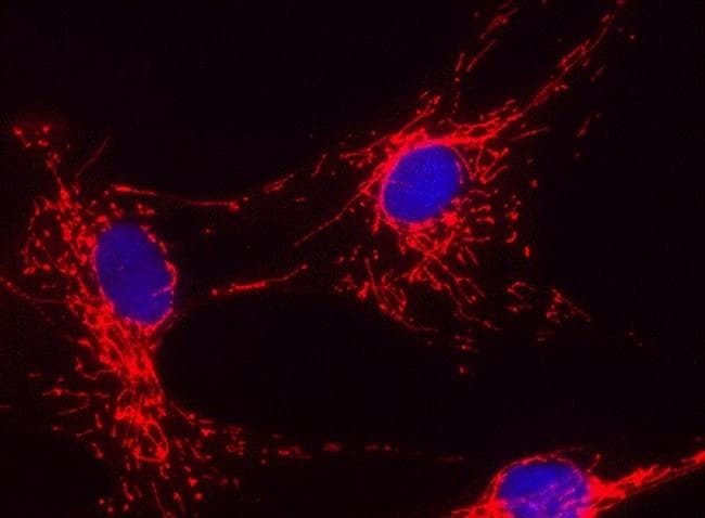
- Immunofluorescent analysis of Cytochrome C in Human fibroblasts using a Cytochrome C Monoclonal antibody (Product # 45-6100) at 2µg/mL. The Human fibroblast cells were fixed and permeabilized, after which they were detected using a fluorescent goat-anti-mouse IgG secondary antibody.

- Submitted by
- Invitrogen Antibodies (provider)
- Main image

- Experimental details
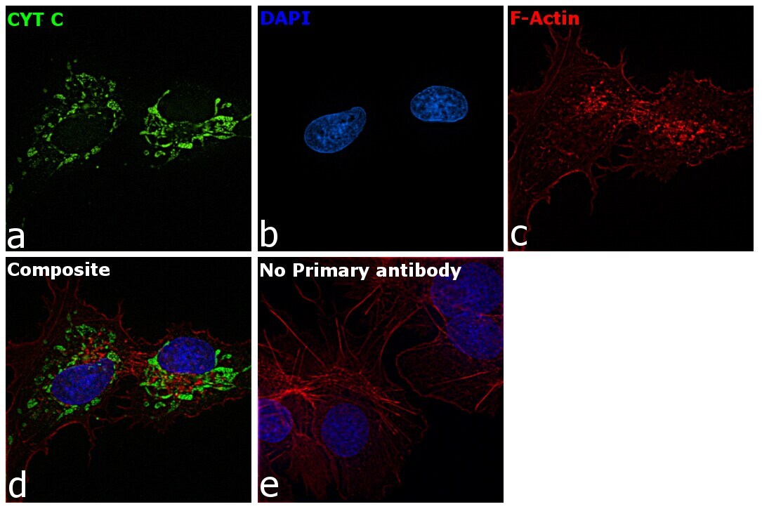
- Immunofluorescence analysis of Cytochrome C was performed using 70% confluent log phase Hep G2 cells. The cells were fixed with 4% paraformaldehyde for 10 minutes, permeabilized with 0.1% Triton™ X-100 for 15 minutes, and blocked with 2% BSA for 45 minutes at room temperature. The cells were labeled with Cytochrome C Monoclonal Antibody (37BA11) (Product # 45-6100) at 1 µg/mL in 0.1% BSA, incubated at 4 degree celsius overnight and then labeled with Donkey anti-Mouse IgG (H+L) Highly Cross-Adsorbed Secondary Antibody, Alexa Fluor Plus 488 (Product # A32766), (1:2000 dilution), for 45 minutes at room temperature (Panel a: Green). Nuclei (Panel b:Blue) were stained with ProLong™ Diamond Antifade Mountant with DAPI (Product # P36962). F-actin (Panel c: Red) was stained with Rhodamine Phalloidin (Product # R415, 1:300 dilution). Panel d represents the merged image showing cytoplasmic (mitochondria-like) localization. Panel e represents control cells with no primary antibody to assess background. The images were captured at 60X magnification.

- Submitted by
- Invitrogen Antibodies (provider)
- Main image

- Experimental details
- Immunofluorescence analysis of Cytochrome C was performed using 70% confluent log phase Hep G2 cells. The cells were fixed with 4% paraformaldehyde for 10 minutes, permeabilized with 0.1% Triton™ X-100 for 15 minutes, and blocked with 2% BSA for 45 minutes at room temperature. The cells were labeled with Cytochrome C Monoclonal Antibody (37BA11) (Product # 45-6100) at 1 µg/mL in 0.1% BSA, incubated at 4 degree celsius overnight and then labeled with Donkey anti-Mouse IgG (H+L) Highly Cross-Adsorbed Secondary Antibody, Alexa Fluor Plus 488 (Product # A32766), (1:2000 dilution), for 45 minutes at room temperature (Panel a: Green). Nuclei (Panel b:Blue) were stained with ProLong™ Diamond Antifade Mountant with DAPI (Product # P36962). F-actin (Panel c: Red) was stained with Rhodamine Phalloidin (Product # R415, 1:300 dilution). Panel d represents the merged image showing cytoplasmic (mitochondria-like) localization. Panel e represents control cells with no primary antibody to assess background. The images were captured at 60X magnification.
- Submitted by
- Invitrogen Antibodies (provider)
- Main image

- Experimental details
- Immunofluorescent analysis of Cytochrome C in Human fibroblasts using a Cytochrome C Monoclonal antibody (Product # 45-6100) at 2µg/mL. The Human fibroblast cells were fixed and permeabilized, after which they were detected using a fluorescent goat-anti-mouse IgG secondary antibody.
Supportive validation
- Submitted by
- Invitrogen Antibodies (provider)
- Main image

- Experimental details
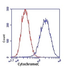
- Flow cytometric analysis of Cytochrome C in HeLa cells using a Cytochrome C Monoclonal antibody (Product # 45-6100) at 1µg/mL, shown in blue. Isotype control antibody shown in red.

- Submitted by
- Invitrogen Antibodies (provider)
- Main image
- Experimental details
- Flow cytometric analysis of Cytochrome C in HeLa cells using a Cytochrome C Monoclonal antibody (Product # 45-6100) at 1µg/mL, shown in blue. Isotype control antibody shown in red.

Supportive validation
- Submitted by
- Invitrogen Antibodies (provider)
- Main image

- Experimental details
- NULL
- Submitted by
- Invitrogen Antibodies (provider)
- Main image

- Experimental details
- NULL
- Submitted by
- Invitrogen Antibodies (provider)
- Main image
- Experimental details
- NULL

- Submitted by
- Invitrogen Antibodies (provider)
- Main image
- Experimental details
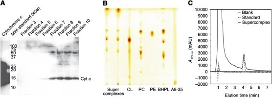
- Figure 5 The supercomplex contains cytochrome c , ubiquinol, and cardiolipin. ( A ) Western blot showing the presence of cytochrome c in the supercomplex. Amphipol-solubilized supercomplex was purified by density gradient centrifugation and fractions were analysed by 15% SDS-PAGE and western blot with an anti-cytochrome c antibody. Cytochrome c runs at ~15 kDa. ( B ) Lipid extracts from two different supercomplex preparations (lanes 1 and 2) and purified lipid standards (lanes 3-5). The supercomplex contains phosphatidyl choline (PC), phosphatidyl ethanolamine (PE), and cardiolipin (CL), which is enriched compared with bovine heart polar lipid extract (BHPL, lane 6). ( C ) Ubiquinol was quantified by HPLC and comparison to Q 10 standards. Each supercomplex contains at least 1 molecule of ubiquinol.
- Submitted by
- Invitrogen Antibodies (provider)
- Main image
- Experimental details
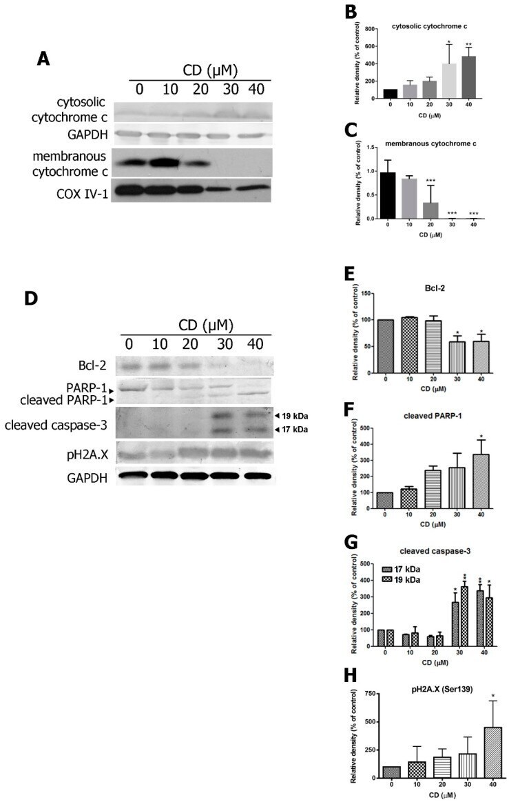
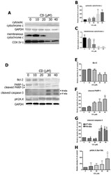
- Figure 5 Effects of CD on pro-apoptotic and anti-apoptotic proteins in T24 BC cells. ( A ) The expression of cytochrome c in the cytosolic fraction and membrane fractions. Glyceraldehyde 3 phosphate dehydrogenase (GAPDH) and cytochrome oxidase subunit IV isoform 1 (COX IV-1) were used as internal controls. ( B , C ) Quantifications of protein expression. ( D ) The expression of Bcl-2, cleaved PARP-1, 17 kDa, and 19 kDa subunits of cleavage caspase-3 and pH2A.X. ( E - H ) Quantifications of the protein expression. Bar graphs show normalised values as percentages relative to the control. Data are expressed as mean +- SD of three to five independent experiments. * p < 0.05, ** p < 0.01, *** p < 0.001 compared with the control group (0 uM).