 Explore
 Validate
 Learn
Learn Western blot
Antibody data
- Antibody Data
- Antigen structure
- References [0]
- Comments [0]
- Validations
- Western blot [1]
- Immunocytochemistry [1]
- Immunohistochemistry [6]
Submit
Validation data
Reference
Comment
Report error
- Product number
- R31747 - Provider product page

- Provider
- NSJ Bioreagents
- Product name
- Cytochrome C Antibody
- Antibody type
- Polyclonal
- Antigen
- Human partial recombinant protein (AA 2-105) was used as the immunogen for this Cytochrome C antibody.
- Description
- Antigen affinity purified antibody
- Reactivity
- Human, Mouse, Rat
- Host
- Rabbit
- Conjugate
- Unconjugated
- Vial size
- 100 µg
- Concentration
- Lyophilized; resuspend with 200 ul for 0.5 mg/ml
- Storage
- Store the Cytochrome C antibody at 4°C. After reconstitution, aliquot and store at -20°C. Avoid repeated freezing and thawing.
No comments: Submit comment
Supportive validation
- Submitted by
- NSJ Bioreagents (provider)
- Main image

- Experimental details
- Western blot testing of Cytochrome C antibody and Lane 1: rat brain; 2: mouse brain; 3: rat heart; 4: mouse heart; 5: U87; 6: Neuro-2a; 7: HeLa; 8: Jurkat; 9: human placenta lysate

Supportive validation
- Submitted by
- NSJ Bioreagents (provider)
- Main image
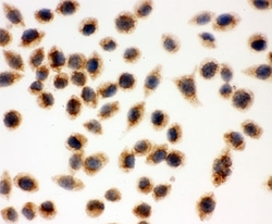
- Experimental details
- ICC testing of Cytochrome C antibody and SMMC-7721 cells

Supportive validation
- Submitted by
- NSJ Bioreagents (provider)
- Main image

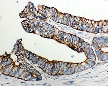
- Experimental details
- IHC-P: Cytochrome C antibody testing of human intestinal cancer tissue. HIER: steamed with pH6 citrate buffer.
- Submitted by
- NSJ Bioreagents (provider)
- Main image
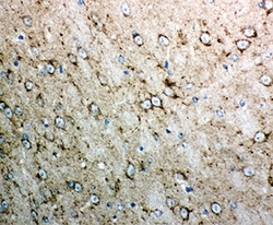
- Experimental details
- IHC-P testing of mouse brain tissue. HIER: steamed with pH6 citrate buffer.

- Submitted by
- NSJ Bioreagents (provider)
- Main image

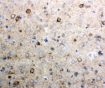
- Experimental details
- IHC-P testing of rat brain tissue. HIER: steamed with pH6 citrate buffer.
- Submitted by
- NSJ Bioreagents (provider)
- Main image

- Experimental details
- Immunofluorescent staining of FFPE human intestinal cancer tissue with Cytochrome C antibody (green) at 1ug/ml and DAPI nuclear stain (blue). HIER: steam section in pH6 citrate buffer for 20 min.

- Submitted by
- NSJ Bioreagents (provider)
- Main image

- Experimental details
- Immunofluorescent staining of FFPE mouse heart tissue with Cytochrome C antibody (green) at 1ug/ml and DAPI nuclear stain (blue). HIER: steam section in pH6 citrate buffer for 20 min.

- Submitted by
- NSJ Bioreagents (provider)
- Main image

- Experimental details
- Immunofluorescent staining of FFPE rat heart tissue with Cytochrome C antibody (green) at 1ug/ml and DAPI nuclear stain (blue). HIER: steam section in pH6 citrate buffer for 20 min.
