Explore
 Validate
 Learn
Learn Flow cytometry
Antibody data
- Antibody Data
- Antigen structure
- References [3]
- Comments [0]
- Validations
- Flow cytometry [1]
- Other assay [2]
Submit
Validation data
Reference
Comment
Report error
- Product number
- 14-6601-82 - Provider product page

- Provider
- Invitrogen Antibodies
- Product name
- Cytochrome C Monoclonal Antibody (6H2), eBioscience™
- Antibody type
- Monoclonal
- Antigen
- Other
- Description
- Description: The 6H2 antibody reacts with the native form of mouse, human, and rat cytochrome c.
- Antibody clone number
- 6H2
- Concentration
- 0.5 mg/mL
Submitted references Metabolite and thymocyte development defects in ADA-SCID mice receiving enzyme replacement therapy.
Caspase dependent programmed cell death in developing embryos: a potential target for therapeutic intervention against pathogenic nematodes.
Common structural features among monoclonal antibodies binding the same antigenic region of cytochrome c.
Moretti FA, Giardino G, Attenborough TCH, Gkazi AS, Margetts BK, la Marca G, Fairbanks L, Crompton T, Gaspar HB
Scientific reports 2021 Dec 1;11(1):23221
Scientific reports 2021 Dec 1;11(1):23221
Caspase dependent programmed cell death in developing embryos: a potential target for therapeutic intervention against pathogenic nematodes.
Mohapatra AD, Kumar S, Satapathy AK, Ravindran B
PLoS neglected tropical diseases 2011 Sep;5(9):e1306
PLoS neglected tropical diseases 2011 Sep;5(9):e1306
Common structural features among monoclonal antibodies binding the same antigenic region of cytochrome c.
Goshorn SC, Retzel E, Jemmerson R
The Journal of biological chemistry 1991 Feb 5;266(4):2134-42
The Journal of biological chemistry 1991 Feb 5;266(4):2134-42
No comments: Submit comment
Supportive validation
- Submitted by
- Invitrogen Antibodies (provider)
- Main image
- Experimental details
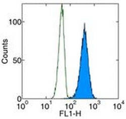
- Intracellular staining of HeLa cells with Anti-Cytochrome C FITC. Appropriate isotype controls were used (open histogram). Total cells were used for analysis.

Supportive validation
- Submitted by
- Invitrogen Antibodies (provider)
- Main image
- Experimental details
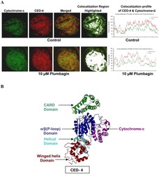
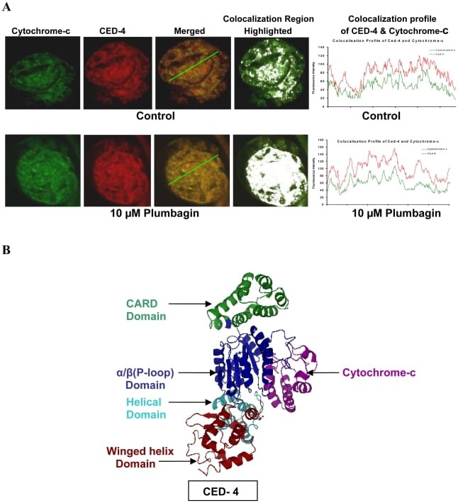
- Figure 5 Demonstration of Cytochrome-c-CED-4 interaction in S.digitata and docking of Cytochrome-c and CED-4 of C.elegans. [ A ] Confocal images of untreated control [upper panel] or Plumbagin treated [lower panel] late embryonic stages demonstrating enhanced colocalization of Cytochrome-c and CED-4 after 24 hr Plumbagin treatment are shown. Regions of colocalization are highlighted as white patches using Image J software. Fluorescence intensity line profile/colocalization profile analysis for both the labeled proteins-Cytochrome-c and CED-4 in control as well as apoptotic embryos, revealing enhanced cololocalization of these proteins in the later are shown [ B ] Interaction of Cytochrome-c of C.elegans [ Magenta ] with alpha/beta [P-loop] ATP binding domain [ Blue ] of CED-4 is shown.
- Submitted by
- Invitrogen Antibodies (provider)
- Main image
- Experimental details
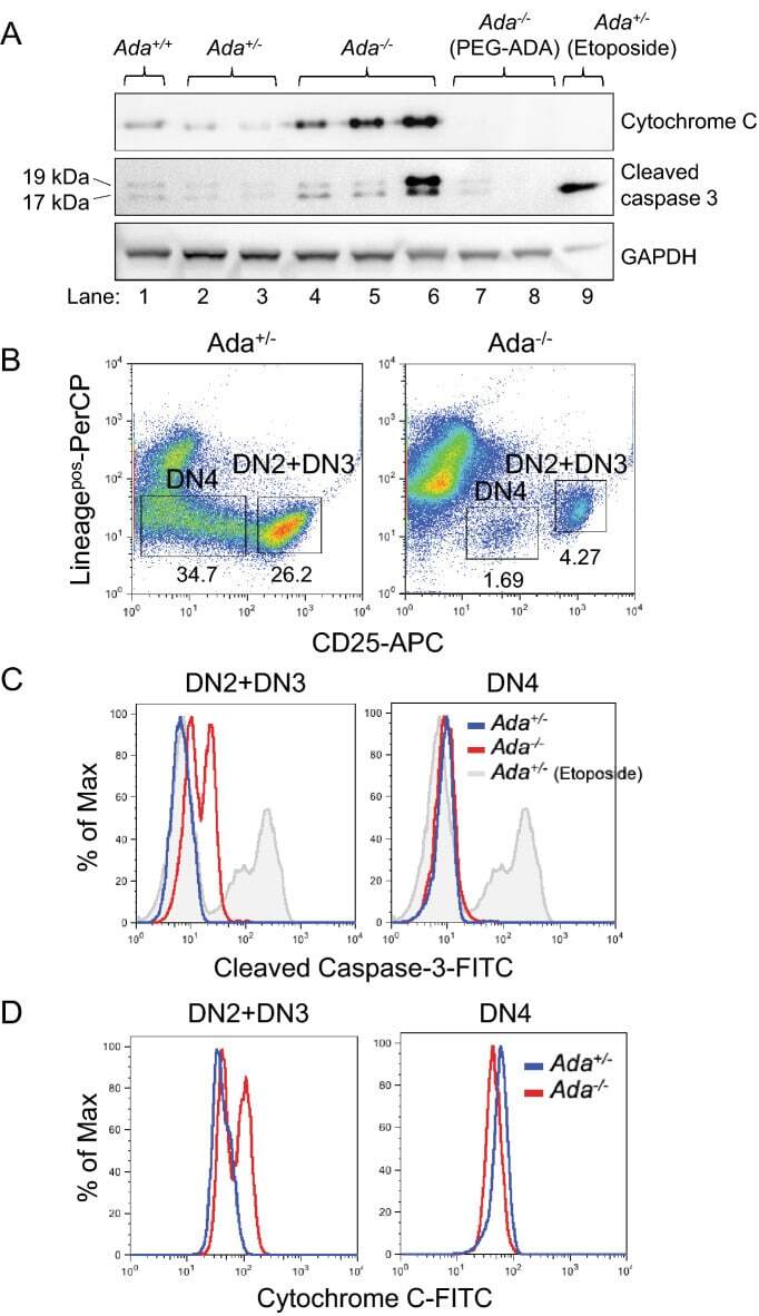
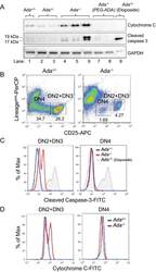
- Figure 3 Immature thymocytes from ADA-deficient mice show increased expression of apoptotic markers. ( A ) Western blot analysis of protein lysates of thymi from P14 untreated mice (lane 1-6) and 3-month-PEG-ADA-treated mice (lane 7-8). As positive control for cleaved caspase-3 staining, unfractionated thymocytes were stimulated in vitro with the apoptosis-inducer etoposide (line 9). Anti-GAPDH stain was used as protein loading control. Full-length blots are presented in Supplementary Fig. 11 . ( B ) FACS analysis of MACS-enriched Lin neg thymocytes from untreated mice at P14. DN4 (Lin neg , CD25 - ) and DN2 + DN3 (Lin neg , CD25 + ) populations are gated. ( C ) Intracellular staining for cleaved caspase-3 of cell populations shown in (B). Unfractionated Lin neg thymocytes were stimulated in vitro with etoposide as apoptosis positive control. ( D ) Intracellular staining for cytochrome C of cell populations shown in (B). FACS plots in (C) and (D) are representative of two replicate experiments (n = 4, 2).