 Explore
 Validate
 Learn
Learn Western blot
Antibody data
- Antibody Data
- Antigen structure
- References [0]
- Comments [0]
- Validations
- Western blot [1]
- Immunohistochemistry [1]
Submit
Validation data
Reference
Comment
Report error
- Product number
- PA3-045 - Provider product page

- Provider
- Invitrogen Antibodies
- Product name
- GPR101 Polyclonal Antibody
- Antibody type
- Polyclonal
- Antigen
- Other
- Description
- Applications Tested: This HTA125 antibody has been pre-diluted and tested by flow cytometric analysis of human TLR4/MD2-transfected cells. This may be used at 5 µL (0.5 µg) per test. A test is defined as the amount (µg) of antibody that will stain a cell sample in a final volume of 100 µL. Cell number should be determined empirically but can range from 10^5 to 10^8 cells/test.
- Reactivity
- Human
- Host
- Rabbit
- Isotype
- IgG
- Vial size
- 100 µL
- Concentration
- Conc. Not Determined
- Storage
- -20° C, Avoid Freeze/Thaw Cycles
No comments: Submit comment
Supportive validation
- Submitted by
- Invitrogen Antibodies (provider)
- Main image

- Experimental details
- Western blot analysis of GPR101 was performed by loading equal amounts of wheat germ lectin agarose bead enriched GPR receptor fractions from mock-transfected or GPR101 transfected HEK293 lysates onto a 7.5% Tris-HCl polyacrylamide gel. Proteins were transferred to a PVDF membrane, blocked and probed with a GPR101 polyclonal antibody (Product # PA3-045) at a dilution of 1:5000, overnight at 4C on a rocking platform, followed by an HRP-conjugated goat anti-rabbit IgG secondary antibody. Denatured GPR101 was detected at ~64kDa. Chemiluminescent detection was performed using ECL.

Supportive validation
- Submitted by
- Invitrogen Antibodies (provider)
- Main image
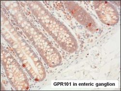
- Experimental details
- Immunohistochemistry analysis of GPR101 was performed on enteric ganglion tissue. To expose target proteins, antigen retrieval was performed by microwaving tissues for 20 minutes in 10mM sodium citrate buffer (pH 6.0). Tissue slides were probed with a GPR101 polyclonal antibody (Product # PA3-045) at a dilution of 1:3000, overnight at 4C in a humidified chamber. Tissues were washed, and detection was performed using an ABC kit composed of biotinylated goat anti-rabbit IgG, peroxidase-conjugated avidin, and 3-amino-9-ethylcarbazole (AEC) substrate in acetate buffer. Tissues were counterstained with hematoxylin and dehydrated to prep for mounting.
