Explore
 Validate
 Learn
Learn Western blot
Antibody data
- Antibody Data
- Antigen structure
- References [0]
- Comments [0]
- Validations
- Western blot [1]
- Immunocytochemistry [1]
- Proximity ligation assay [3]
Submit
Validation data
Reference
Comment
Report error
- Product number
- H00003055-D01P - Provider product page

- Provider
- Abnova Corporation
- Proper citation
- Abnova Corporation Cat#H00003055-D01P, RRID:AB_1575815
- Product name
- HCK purified MaxPab rabbit polyclonal antibody (D01P)
- Antibody type
- Polyclonal
- Description
- Rabbit polyclonal antibody raised against a full-length human HCK protein.
- Antigen sequence
MGCMKSKFLQVGGNTFSKTETSASPHCPVYVPDPT
STIKPGPNSHNSNTPGIREAGSEDIIVVALYDYEA
IHHEDLSFQKGDQMVVLEESGEWWKARSLATRKEG
YIPSNYVARVDSLETEEWFFKGISRKDAERQLLAP
GNMLGSFMIRDSETTKGSYSLSVRDYDPRQGDTVK
HYKIRTLDNGGFYISPRSTFSTLQELVDHYKKGND
GLCQKLSVPCMSSKPQKPWEKDAWEIPRESLKLEK
KLGAGQFGEVWMATYNKHTKVAVKTMKPGSMSVEA
FLAEANVMKTLQHDKLVKLHAVVTKEPIYIITEFM
AKGSLLDFLKSDEGSKQPLPKLIDFSAQIAEGMAF
IEQRNYIHRDLRAANILVSASLVCKIADFGLARVI
EDNEYTAREGAKFPIKWTAPEAINFGSFTIKSDVW
SFGILLMEIVTYGRIPYPGMSNPEVIRALERGYRM
PRPENCPEELYNIMMRCWKNRPEERPTFEYIQSVL
DDFYTATESQYQQQP- Storage
- Store at -20°C or lower. Aliquot to avoid repeated freezing and thawing.
No comments: Submit comment
Supportive validation
- Submitted by
- Abnova Corporation (provider)
- Main image

- Experimental details
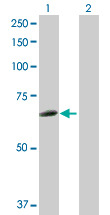
- Western Blot analysis of HCK expression in transfected 293T cell line (H00003055-T02) by HCK MaxPab polyclonal antibody.Lane 1: HCK transfected lysate(57.30 KDa).Lane 2: Non-transfected lysate.
Supportive validation
- Submitted by
- Abnova Corporation (provider)
- Main image
- Experimental details
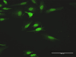
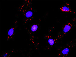
- Immunofluorescence of purified MaxPab antibody to HCK on HeLa cell. [antibody concentration 10 ug/ml]
- Validation comment
- Immunofluorescence
- Protocol
- Protocol

Supportive validation
- Submitted by
- Abnova Corporation (provider)
- Main image
- Experimental details
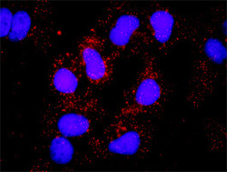
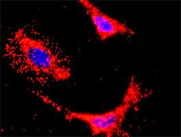
- Proximity Ligation Analysis of protein-protein interactions between HCK and SOS1. Huh7 cells were stained with anti-HCK rabbit purified polyclonal 1:1200 and anti-SOS1 mouse monoclonal antibody 1:50. Each red dot represents the detection of protein-protein interaction complex, and nuclei were counterstained with DAPI (blue).
- Validation comment
- In situ Proximity Ligation Assay (Cell)

- Submitted by
- Abnova Corporation (provider)
- Main image

- Experimental details
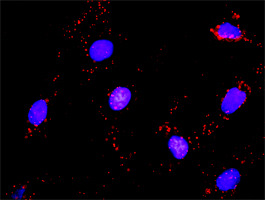
- Proximity Ligation Analysis of protein-protein interactions between HCK and PIK3CB. HeLa cells were stained with anti-HCK rabbit purified polyclonal 1:1200 and anti-PIK3CB mouse monoclonal antibody 1:50. Each red dot represents the detection of protein-protein interaction complex, and nuclei were counterstained with DAPI (blue).
- Validation comment
- In situ Proximity Ligation Assay (Cell)
- Submitted by
- Abnova Corporation (provider)
- Main image
- Experimental details
- Proximity Ligation Analysis of protein-protein interactions between HCK and CRKL. Mahlavu cells were stained with anti-HCK rabbit purified polyclonal 1:1200 and anti-CRKL mouse monoclonal antibody 1:50. Each red dot represents the detection of protein-protein interaction complex, and nuclei were counterstained with DAPI (blue).
- Validation comment
- In situ Proximity Ligation Assay (Cell)