 Explore
 Validate
 Learn
Learn Western blot
Antibody data
- Antibody Data
- Antigen structure
- References [0]
- Comments [0]
- Validations
- Western blot [2]
- Immunocytochemistry [1]
Submit
Validation data
Reference
Comment
Report error
- Product number
- Ab106982 - Provider product page

- Provider
- Aladdin Scientific
- Product name
- Hck Mouse mAb
- Antibody type
- Monoclonal
- Description
- Mouse anti Human Hck Antibody, Monoclonal (1508CT602.13.1), could be used for WB, ICC, IF and so on.ApplicationWB: 1/2000-1/4000ICC/IF: 1/25Protein FunctionMay serve as part of a signaling pathway coupling the Fc receptor to the activation of the respiratory burst. May also contribute to neutrophil migration and may regulate the degranulation process of neutrophils.
- Reactivity
- Human, Mouse, Rat
- Host
- Mouse
- Conjugate
- Unconjugated
- Antigen sequence
AA 1-526- Antibody clone number
- 1508CT602.13.1
- Vial size
- 100μl,10μl,1ml,50μl
- Concentration
- 0,5 mg/ml
- Storage
- Store at 4℃ short term (1-2 weeks). Store at -20℃ long term (24 months). Upon delivery aliquot. Avoid freeze/thaw cycle.
No comments: Submit comment
Supportive validation
- Submitted by
- Aladdin Scientific (provider)
- Main image
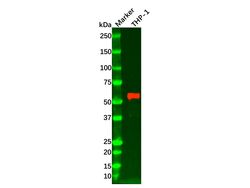
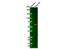
- Experimental details
- Hck Mouse mAb (Ab106982) - Western Blot All lanes: Hck Mouse mAb (Ab106982) at 1/2000 dilution Samples: Lysates at 20 µg per lane Secondary: Goat Anti-Mouse IgG H&L (HRP) (Ab138040) at 1/30000 dilution Predicted band size: 60 kDa Observed band size: 58 kDa Exposure time: 10.0 s
- Submitted by
- Aladdin Scientific (provider)
- Main image
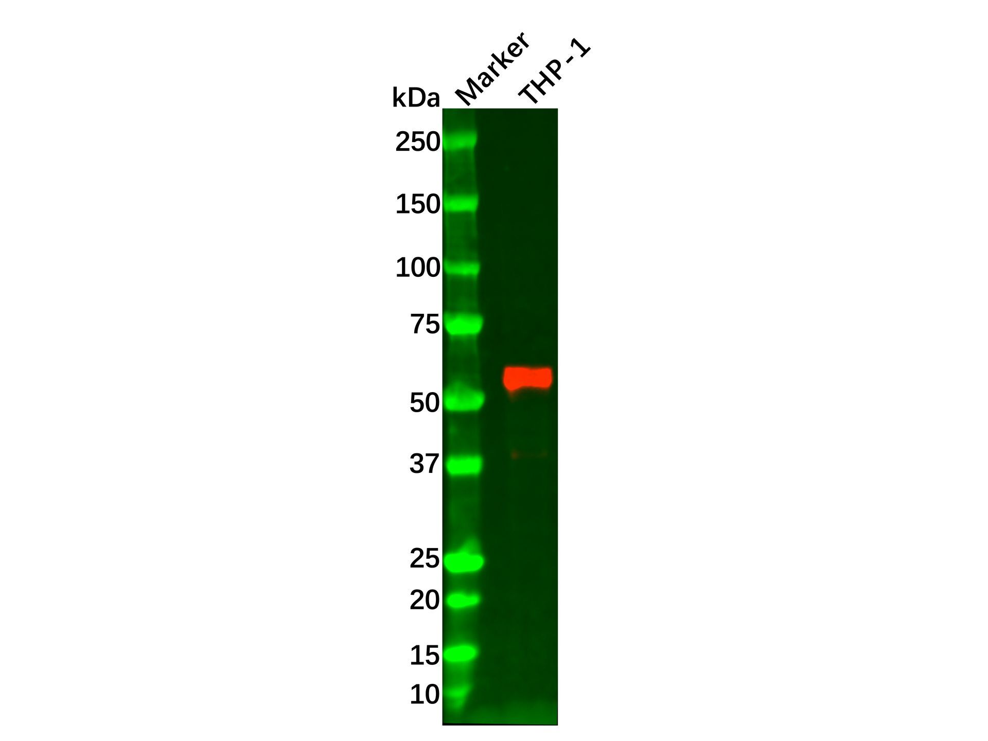
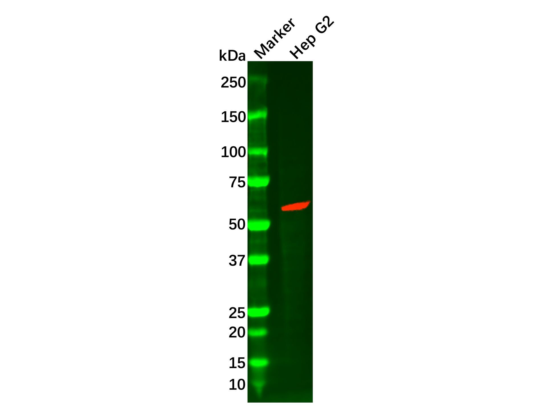
- Experimental details
- Hck Mouse mAb (Ab106982) - Western Blot All lanes: Hck Mouse mAb (Ab106982) at 1/2000 dilution Samples: Lysates at 20 µg per lane Secondary: Goat Anti-Mouse IgG H&L (HRP) (Ab138040) at 1/30000 dilution Predicted band size: 60 kDa Observed band size: 58 kDa Exposure time: 60.0 s
Supportive validation
- Submitted by
- Aladdin Scientific (provider)
- Main image
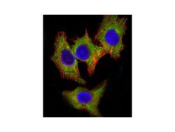
- Experimental details
- Hck Mouse mAb (Ab106982) - IF/ICC Immunofluorescent analysis of 4% paraformaldehyde-fixed, 0.1% Triton X-100 permeabilized Hep G2 (human liver hepatocellular carcinoma cell line) cells labeling HCK with Hck Mouse mAb (Ab106982) at 1/25 dilution, followed by Dylight® 488-conjugated goat anti-mouse IgG secondary antibody at 1/200 dilution (green). Immunofluorescence image showing cytoplasm staining on HepG2 cell line. Cytoplasmic actin is detected with Dylight® 554 Phalloidin at 1/100 dilution (red). The nuclear counter stain is DAPI (blue).
