Explore
 Validate
 Learn
Learn Western blot
 Immunocytochemistry
Antibody data
- Antibody Data
- Antigen structure
- References [1]
- Comments [0]
- Validations
- Immunocytochemistry [1]
- Immunohistochemistry [1]
- Other assay [1]
Submit
Validation data
Reference
Comment
Report error
- Product number
- PA5-105531 - Provider product page

- Provider
- Invitrogen Antibodies
- Product name
- Phospho-FGFR4 (Tyr642) Polyclonal Antibody
- Antibody type
- Polyclonal
- Antigen
- Synthetic peptide
- Description
- Antibody detects endogenous levels of FGFR4 only when phosphorylated at Tyr642.
- Reactivity
- Human, Mouse, Rat
- Host
- Rabbit
- Isotype
- IgG
- Vial size
- 100 μL
- Concentration
- 1 mg/mL
- Storage
- -20°C
Submitted references Gait disturbances and muscle dysfunction in fibroblast growth factor 2 knockout mice.
Homer-Bouthiette C, Xiao L, Hurley MM
Scientific reports 2021 May 26;11(1):11005
Scientific reports 2021 May 26;11(1):11005
No comments: Submit comment
Supportive validation
- Submitted by
- Invitrogen Antibodies (provider)
- Main image
- Experimental details
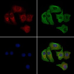
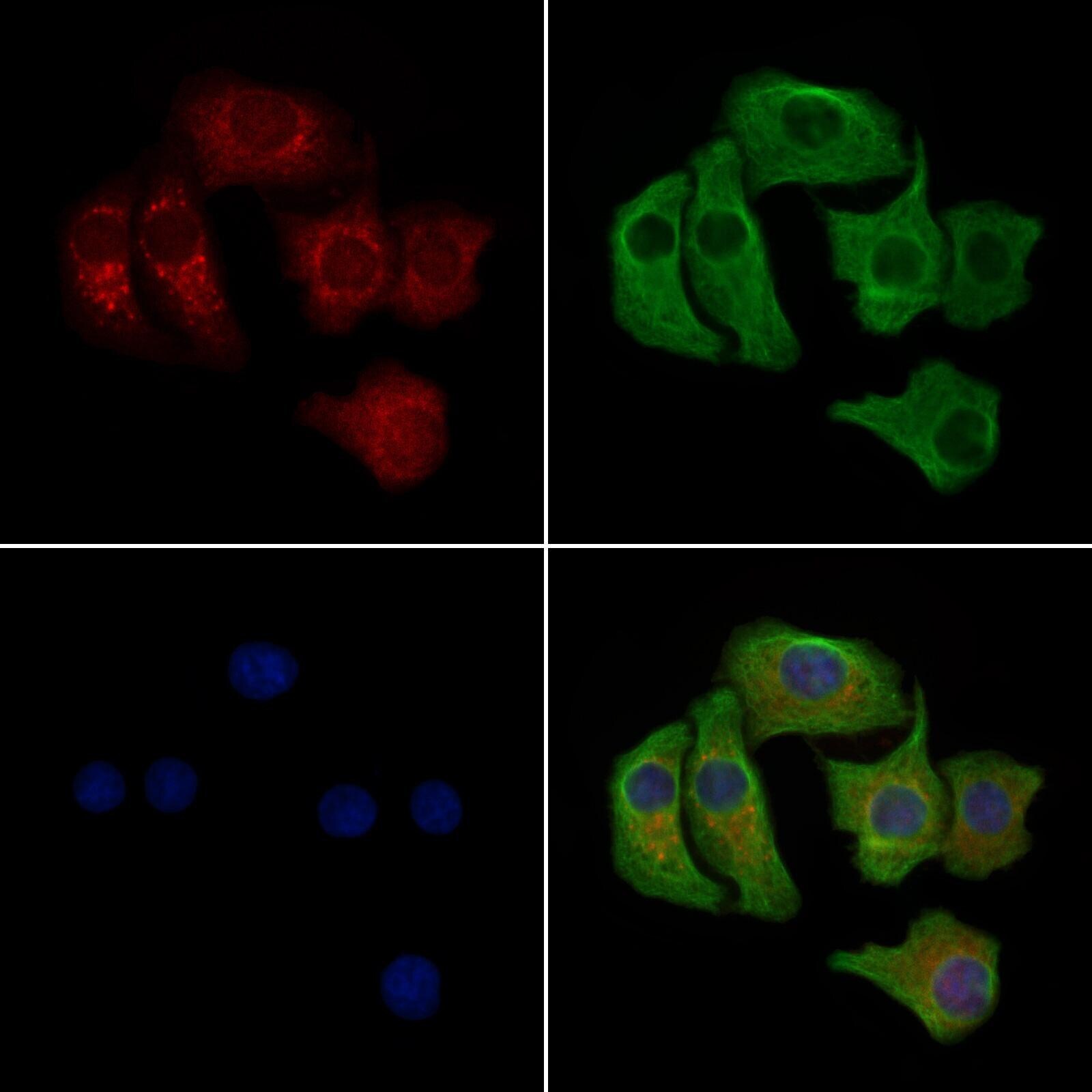
- Immunocytochemistry analysis of Phospho-FGFR4 (Tyr642) in Hela cells (heat shock treatment). The samples were fixed with PFA and permeabilized in 0.1% Triton X-100, then blocked in 10% serum for 45 minutes at 25°C. Samples were incubated with Phospho-FGFR4 (Tyr642) polyclonal antibody (Product # PA5-105531) and mouse anti-beta tubulin antibody for 1 hour at 37°C. An AlexaFluor594 conjugated goat anti-rabbit IgG(H+L) (Red) and an AlexaFluor488 conjugated goat anti-mouse IgG(H+L) (Green) were used as the secondary antibodies. The nuclear counter stain is DAPI (blue).
Supportive validation
- Submitted by
- Invitrogen Antibodies (provider)
- Main image
- Experimental details
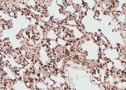
- Immunohistochemistry analysis of paraffin-embedded Phospho-FGFR4 (Tyr642) in rat lung tissue sections. Antigen retrieval was performed using citrate buffer. Samples were blocked with blocking buffer (1.5 hr, 22°C), incubated with Phospho-FGFR4 (Tyr642) polyclonal antibody (Product # PA5-105531) using a dilution of 1:100 (1.5 hr, 22°C), followed by HRP conjugated goat anti-rabbit.

Supportive validation
- Submitted by
- Invitrogen Antibodies (provider)
- Main image
- Experimental details
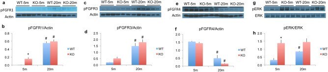
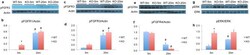
- Figure 8 Expression of FGFR protein and MAPK/ERK signaling in skeletal muscle from WT and Fgf2KO mice. Young (5 m) and old (20 m) WT and Fgf2KO mouse triceps muscles were isolated and total protein extracted for western blotting. ( a ) phosphoFGFR1 Western blot and ( b ) quantification. ( c ) phosphoFGFR3 Western blot and ( d ) quantification. ( e ) phosphoFGFR4 Western blot and ( f ) quantification. ( g ) phosphoERK and total ERK Western blot and ( h ) quantification . ( a , b ) phosphoFGFR1; ( c , d ) phosphoFGFR3 were significantly increased, while ( e , f ) phosphoFGFR4 was significantly decreased in old muscle of both genotypes. ( g , h ) phosphoERK1/2 was significantly increased in 20 m old as well 5 m and 20 m Fgf2KO compared with 5 m WT. n = 3 mice/group. *Compared to corresponding WT p < 0.05, # compared to corresponding 5 m p < 0.05.