Explore
 Validate
 Learn
Learn Western blot
Antibody data
- Antibody Data
- Antigen structure
- References [0]
- Comments [0]
- Validations
- Western blot [2]
- Immunocytochemistry [2]
- Immunohistochemistry [1]
- Flow cytometry [1]
Submit
Validation data
Reference
Comment
Report error
- Product number
- F50850 - Provider product page

- Provider
- NSJ Bioreagents
- Product name
- Gapdh Antibody
- Antibody type
- Polyclonal
No comments: Submit comment
Supportive validation
- Submitted by
- NSJ Bioreagents (provider)
- Main image
- Experimental details
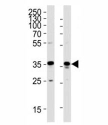
- Western blot analysis of lysate from HeLa, HUVEC cell line (left to right) using GAPDH antibody; Ab was diluted at 1:1000 for each lane.

- Submitted by
- NSJ Bioreagents (provider)
- Main image

- Experimental details
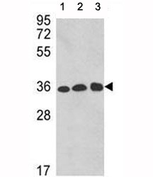
- Western blot analysis of GAPDH antibody and 1) A2058, 2) A375, and 3) CEM lysate
Supportive validation
- Submitted by
- NSJ Bioreagents (provider)
- Main image
- Experimental details
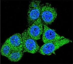
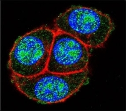
- Confocal immunofluorescent analysis of GAPDH antibody with HeLa cells followed by Alexa Fluor 488-conjugated goat anti-rabbit lgG (green). DAPI was used as a nuclear counterstain (blue).

- Submitted by
- NSJ Bioreagents (provider)
- Main image
- Experimental details
- Confocal immunofluorescent analysis of GAPDH antibody with HeLa cells followed by Alexa Fluor 488-conjugated goat anti-rabbit lgG (green). Actin filaments have been labeled with Alexa Fluor 555 Phalloidin (red). DAPI was used as a nuclear counterstain (blue).

Supportive validation
- Submitted by
- NSJ Bioreagents (provider)
- Main image

- Experimental details
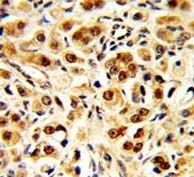
- IHC analysis of FFPE human prostate carcinoma with GAPDH antibody
Supportive validation
- Submitted by
- NSJ Bioreagents (provider)
- Main image

- Experimental details
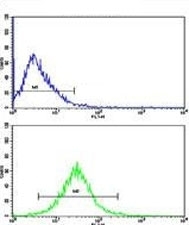
- Flow cytometric analysis of HepG2 cells using GAPDH antibody (bottom histogram) compared to a negative control cell (top histogram). FITC-conjugated goat-anti-rabbit secondary Ab was used for the analysis.