Explore
 Validate
 Learn
Learn Western blot
 Immunocytochemistry
 Immunohistochemistry
Antibody data
- Antibody Data
- Antigen structure
- References [5]
- Comments [0]
- Validations
- Western blot [1]
- Immunocytochemistry [1]
Submit
Validation data
Reference
Comment
Report error
- Product number
- AMAb91153 - Provider product page

- Provider
- Atlas Antibodies
- Proper citation
- Atlas Antibodies Cat#AMAb91153, RRID:AB_2665822
- Product name
- Anti-GAPDH
- Antibody type
- Monoclonal
- Description
- Monoclonal Antibody against Human GAPDH, Clone ID: CL3266, Gene description: glyceraldehyde-3-phosphate dehydrogenase, Alternative Gene Names: GAPD, Validated applications: IHC, WB, ICC, Uniprot ID: P04406, Storage: Store at +4°C for short term storage. Long time storage is recommended at -20°C.
- Reactivity
- Human
- Host
- Mouse
- Conjugate
- Unconjugated
- Isotype
- IgG
- Antibody clone number
- CL3266
- Vial size
- 100 µl
- Concentration
- 1.0 mg/ml
- Storage
- Store at +4°C for short term storage. Long time storage is recommended at -20°C.
- Handling
- The antibody solution should be gently mixed before use.
Submitted references Pomegranate Extract Affects Gut Biofilm Forming Bacteria and Promotes Intestinal Mucosal Healing Regulating the Crosstalk between Epithelial Cells and Intestinal Fibroblasts
ZDHHC9 promotes colon tumor growth by inhibiting effector T cells
ADNCR modulates neural stem cell differentiation and proliferation through the regulation of TCF3 expression
Long non‑coding RNA SNHG1 promotes breast cancer progression by regulation of LMO4
Long Stress Induced Non-Coding Transcripts 5 (LSINCT5) Promotes Hepatocellular Carcinoma Progression Through Interaction with High-Mobility Group AT-hook 2 and MiR-4516
Rizzo G, Pineda Chavez S, Vandenkoornhuyse E, Cárdenas Rincón C, Cento V, Garlatti V, Wozny M, Sammarco G, Di Claudio A, Meanti L, Elangovan S, Romano A, Roda G, Loy L, Dal Buono A, Gabbiadini R, Lovisa S, Rusconi R, Repici A, Armuzzi A, Vetrano S
Nutrients 2023;15(7):1771
Nutrients 2023;15(7):1771
ZDHHC9 promotes colon tumor growth by inhibiting effector T cells
Chong X, Zhu L, Yu D, Chen S, Wang G, Yu Q, Ma X, Xu J, Chen H, An H
Oncology Letters 2022;25(1)
Oncology Letters 2022;25(1)
ADNCR modulates neural stem cell differentiation and proliferation through the regulation of TCF3 expression
Long L, Zeng C, Chen H, Zhou T, Wu L, Cai X
Annals of Translational Medicine 2020;8(15):927-927
Annals of Translational Medicine 2020;8(15):927-927
Long non‑coding RNA SNHG1 promotes breast cancer progression by regulation of LMO4
Xiong X, Feng Y, Li L, Yao J, Zhou M, Zhao P, Huang F, Zeng L, Yuan L
Oncology Reports 2020
Oncology Reports 2020
Long Stress Induced Non-Coding Transcripts 5 (LSINCT5) Promotes Hepatocellular Carcinoma Progression Through Interaction with High-Mobility Group AT-hook 2 and MiR-4516
Li O, Li Z, Tang Q, Li Y, Yuan S, Shen Y, Zhang Z, Li N, Chu K, Lei G
Medical Science Monitor 2018;24
Medical Science Monitor 2018;24
No comments: Submit comment
Enhanced validation
- Submitted by
- Atlas Antibodies (provider)
- Enhanced method
- Genetic validation
- Main image
- Experimental details
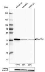
- Western blot analysis in U-251MG cells transfected with control siRNA, target specific siRNA probe #1 and #2, using Anti-GAPDH antibody. Remaining relative intensity is presented. Loading control: Anti-PPIB.
- Sample type
- Human
- Protocol
- Protocol

Supportive validation
- Submitted by
- Atlas Antibodies (provider)
- Main image

- Experimental details
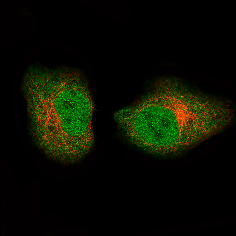
- Immunofluorescence staining of A549 cells using the Anti-GAPDH monoclonal antibody, showing specific staining in the nucleoplasm and cytosol in green. Microtubule- and nuclear probes are visualized in red and blue, respectively (where available).
- Sample type
- Human