Explore
 Validate
 Learn
Learn Western blot
 ELISA
ELISAAntibody data
- Antibody Data
- Antigen structure
- References [5]
- Comments [0]
- Validations
- Western blot [2]
- Immunocytochemistry [1]
- Immunohistochemistry [1]
Submit
Validation data
Reference
Comment
Report error
- Product number
- AP16240PU-T - Provider product page

- Provider
- Acris Antibodies GmbH
- Proper citation
- Acris Antibodies GmbH Cat#AP16240PU-T, RRID:AB_11001859
- Product name
- anti GAPDH (C-term)
- Antibody type
- Polyclonal
- Antigen
- Peptide with sequence from the C-Terminus of the protein sequence according to NP_002037.2.
- Reactivity
- Human, Mouse, Rat, Canine
- Host
- Goat
- Vial size
- 20 µg
- Concentration
- 0.5 mg/ml
Submitted references Distinct molecular mechanisms of acquired resistance to temozolomide in glioblastoma cells.
Defined carboxy-terminal fragments of insulin-like growth factor (IGF) binding protein-2 exert similar mitogenic activity on cultured rat growth plate chondrocytes as IGF-I.
Developmental regulation of the NMDA receptor subunits, NR3A and NR1, in human prefrontal cortex.
Brain fatty acid binding protein (Fabp7) is diurnally regulated in astrocytes and hippocampal granule cell precursors in adult rodent brain.
Mice lacking NKCC1 are protected from development of bacteremia and hypothermic sepsis secondary to bacterial pneumonia.
Happold C, Roth P, Wick W, Schmidt N, Florea AM, Silginer M, Reifenberger G, Weller M
Journal of neurochemistry 2012 Jul;122(2):444-55
Journal of neurochemistry 2012 Jul;122(2):444-55
Defined carboxy-terminal fragments of insulin-like growth factor (IGF) binding protein-2 exert similar mitogenic activity on cultured rat growth plate chondrocytes as IGF-I.
Kiepe D, Van Der Pas A, Ciarmatori S, Ständker L, Schütt B, Hoeflich A, Hügel U, Oh J, Tönshoff B
Endocrinology 2008 Oct;149(10):4901-11
Endocrinology 2008 Oct;149(10):4901-11
Developmental regulation of the NMDA receptor subunits, NR3A and NR1, in human prefrontal cortex.
Henson MA, Roberts AC, Salimi K, Vadlamudi S, Hamer RM, Gilmore JH, Jarskog LF, Philpot BD
Cerebral cortex (New York, N.Y. : 1991) 2008 Nov;18(11):2560-73
Cerebral cortex (New York, N.Y. : 1991) 2008 Nov;18(11):2560-73
Brain fatty acid binding protein (Fabp7) is diurnally regulated in astrocytes and hippocampal granule cell precursors in adult rodent brain.
Gerstner JR, Bremer QZ, Vander Heyden WM, Lavaute TM, Yin JC, Landry CF
PloS one 2008 Feb 20;3(2):e1631
PloS one 2008 Feb 20;3(2):e1631
Mice lacking NKCC1 are protected from development of bacteremia and hypothermic sepsis secondary to bacterial pneumonia.
Nguyen M, Pace AJ, Koller BH
The Journal of experimental medicine 2007 Jun 11;204(6):1383-93
The Journal of experimental medicine 2007 Jun 11;204(6):1383-93
No comments: Submit comment
Supportive validation
- Submitted by
- Acris Antibodies GmbH (provider)
- Main image
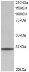
- Experimental details
- AP16240PU-N GAPDH Antibody staining of HEK293 lysate at 0.01 µg/ml (RIPA buffer, 35 µg total protein per lane). Primary incubated for 1 hour. Detected by western blot using Chemiluminescence.
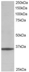
- Submitted by
- Acris Antibodies GmbH (provider)
- Main image
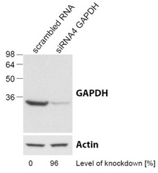
- Experimental details
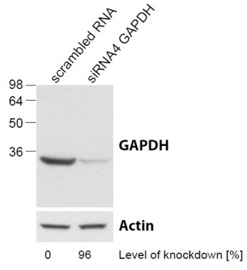
- AP16240PU-N GAPDH Antibody (0.1 µg/ml) staining of HeLa lysate (control in left lane and after si-RNA-mediated GAPDH knock-down expresson in right lane) (35µg protein in RIPA buffer). Level of knock-down relative to Actin expression level was determined by RT-PCR. Primary incubation was 1 hour. Detected by chemiluminescence.
Supportive validation
- Submitted by
- Acris Antibodies GmbH (provider)
- Main image
- Experimental details
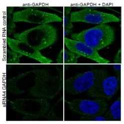
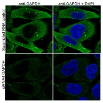
- AP16240PU-N GAPDH Antibody (0.1 µg/ml) staining of PFA-fixed HeLa before (top) and after (bottom) si-RNA-mediated GAPDH knock-down expresson. Primary incubation 1h at ambient temp. Detection by DyLight 488. Nuclear DAPI stain (right).
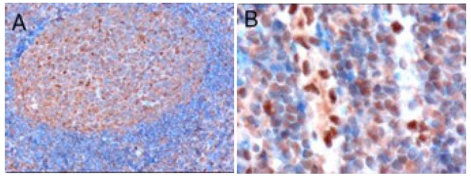
Supportive validation
- Submitted by
- Acris Antibodies GmbH (provider)
- Main image
- Experimental details
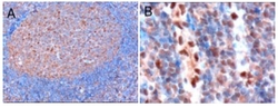
- AP16240PU-N GAPDH Antibody staining of Paraffin Embedded Human Tonsil at 0.3 µg/ml. Microwaved antigen retrieval with Tris/EDTA buffer pH9, HRP-staining. A) Staining of germinal centre cells. B) Staining of endothelial cells.