Explore
 Validate
 Learn
Learn Western blot
 Immunocytochemistry
Antibody data
- Antibody Data
- Antigen structure
- References [1]
- Comments [0]
- Validations
- Immunocytochemistry [1]
- Other assay [1]
Submit
Validation data
Reference
Comment
Report error
- Product number
- 711074 - Provider product page

- Provider
- Invitrogen Antibodies
- Product name
- GDNF Recombinant Superclonal™ Antibody (13HCLC)
- Antibody type
- Other
- Antigen
- Other
- Description
- This antibody is predicted to react with Monkey, Horse and Pig Recombinant rabbit Superclonal™ antibodies are unique offerings from Thermo Fisher Scientific. They are comprised of a selection of multiple different recombinant monoclonal antibodies, providing the best of both worlds - the sensitivity of polyclonal antibodies with the specificity of monoclonal antibodies - all delivered with the consistency only found in a recombinant antibody. While functionally the same as a polyclonal antibody - recognizing multiple epitope sites on the target and producing higher detection sensitivity for low abundance targets - a recombinant rabbit Superclonal™ antibody has a known mixture of light and heavy chains. The exact population can be produced in every lot, circumventing the biological variability typically associated with polyclonal antibody production. Note: Formerly called “Recombinant polyclonal antibody”, this product is now rebranded as “Recombinant Superclonal™ antibody”. The physical product and the performance remain unchanged.
- Reactivity
- Human
- Host
- Rabbit
- Isotype
- IgG
- Antibody clone number
- 13HCLC
- Vial size
- 100 μg
- Concentration
- 0.5 mg/mL
- Storage
- Store at 4°C short term. For long term storage, store at -20°C, avoiding freeze/thaw cycles.
Submitted references In vitro evaluation of periapical lesion-derived stem cells for dental pulp tissue engineering.
Li W, Mao M, Hu N, Wang J, Huang J, Gu S
FEBS open bio 2022 Jan;12(1):270-284
FEBS open bio 2022 Jan;12(1):270-284
No comments: Submit comment
Supportive validation
- Submitted by
- Invitrogen Antibodies (provider)
- Main image
- Experimental details
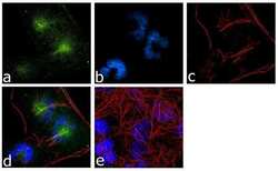
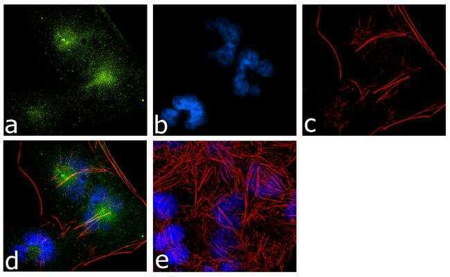
- For immunofluorescence analysis, T98G glioblastoma cells were fixed and permeabilized for detection of endogenous GDNF using Anti- GDNF Recombinant Rabbit Superclonal™ Antibody (Product # 711074, 2 µg/mL) and labeled with Goat anti-Rabbit IgG (Heavy Chain) Superclonal Secondary Antibody, Alexa Fluor® 488 conjugate (Product # A27034, 1:2000). Panel a) shows representative cells that were stained for detection and localization of GDNF protein (green), Panel b) is stained for nuclei (blue) using SlowFade® Gold Antifade Mountant with DAPI (Product # S36938). Panel c) represents cytoskeletal F-actin staining using Alexa Fluor® 555 Rhodamine Phalloidin (Product # R415, 1:300). Panel d) is a composite image of Panels a, b and c clearly demonstrating localization of GDNF in the cytoplasm. Panel e) represents control cells with no primary antibody to assess background. The images were captured at 60X magnification.
Supportive validation
- Submitted by
- Invitrogen Antibodies (provider)
- Main image
- Experimental details
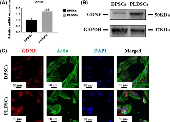
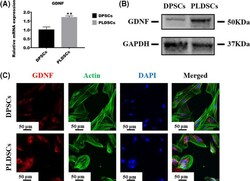
- Fig. 5 Comparison of neurotrophic ability of DPSCs and PLDSCs. (A) qRT-PCR was used to compare the expression level of GDNF between PLDSCs and DPSCs; the expression level was normalized to that of ACTB ( n = 3, Student's t -test). (B) Western blotting results of GDNF expression. (C) Immunofluorescence staining for GDNF expression in DPSCs and PLDSCs. Scale bar = 50 mum. ** P < 0.01. Data are shown as the mean +- SD.