Explore
 Validate
 Learn
Learn Western blot
 Immunocytochemistry
Antibody data
- Antibody Data
- Antigen structure
- References [0]
- Comments [0]
- Validations
- Immunocytochemistry [4]
Submit
Validation data
Reference
Comment
Report error
- Product number
- MA5-45279 - Provider product page

- Provider
- Invitrogen Antibodies
- Product name
- GRP78 Monoclonal Antibody (1H11-1H7), PE
- Antibody type
- Monoclonal
- Antigen
- Other
- Description
- 0.5 µg/mL of MA5-45279 was sufficient for detection of Grp78 in 10 µg of rat tissue lysate by ECL immunoblot analysis.|Detects approximately 78kDa.
- Reactivity
- Human, Mouse, Rat, Bovine, Hamster, Rabbit, Xenopus
- Host
- Mouse
- Conjugate
- Yellow dye
- Isotype
- IgG
- Antibody clone number
- 1H11-1H7
- Vial size
- 100 μg
- Concentration
- 1 mg/mL
- Storage
- 4°C
No comments: Submit comment
Supportive validation
- Submitted by
- Invitrogen Antibodies (provider)
- Main image
- Experimental details
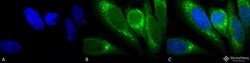
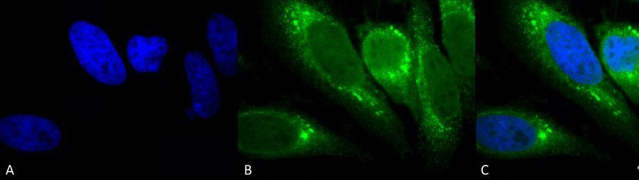
- Immunocytochemistry/Immunofluorescence analysis using human heat shocked cervical cancer cells. Fixation involved 2% Formaldehyde for 20 min at RT. Samples were incubated with GRP78 monoclonal antibody (Product # MA5-45279) at 1:100 for 12 hours at 4°C, followed by FITC Goat Anti-Mouse (green) at 1:200 for 2 hours at RT. Counterstain used was DAPI (blue) nuclear stain at 1:40,000 for 2 hours at RT. Localization: Endoplasmic reticulum lumen. Melanosome. Magnification: 100x. (A) DAPI (blue) nuclear stain. (B) Anti-GRP78 antibody. (C) Composite. Heat Shocked at 42°C for 1h.
- Conjugate
- Yellow dye
- Submitted by
- Invitrogen Antibodies (provider)
- Main image
- Experimental details
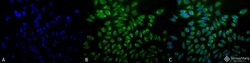
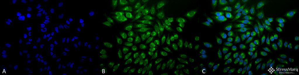
- Immunocytochemistry/Immunofluorescence analysis using human heat shocked cervical cancer cells. Fixation involved 2% Formaldehyde for 20 min at RT. Samples were incubated with GRP78 monoclonal antibody (Product # MA5-45279) at 1:100 for 12 hours at 4°C, followed by FITC Goat Anti-Mouse (green) at 1:200 for 2 hours at RT. Counterstain used was DAPI (blue) nuclear stain at 1:40,000 for 2 hours at RT. Localization: Endoplasmic reticulum lumen. Melanosome. Magnification: 20x. (A) DAPI (blue) nuclear stain. (B) Anti-GRP78 antibody. (C) Composite. Heat Shocked at 42°C for 1h.
- Conjugate
- Yellow dye
- Submitted by
- Invitrogen Antibodies (provider)
- Main image
- Experimental details
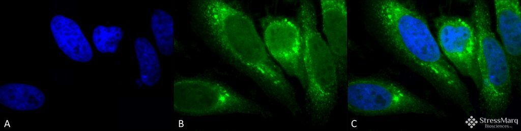
- Immunocytochemistry/Immunofluorescence analysis using human heat shocked cervical cancer cells. Fixation involved 2% Formaldehyde for 20 min at RT. Samples were incubated with GRP78 monoclonal antibody (Product # MA5-45279) at 1:100 for 12 hours at 4°C, followed by FITC Goat Anti-Mouse (green) at 1:200 for 2 hours at RT. Counterstain used was DAPI (blue) nuclear stain at 1:40,000 for 2 hours at RT. Localization: Endoplasmic reticulum lumen. Melanosome. Magnification: 100x. (A) DAPI (blue) nuclear stain. (B) Anti-GRP78 antibody. (C) Composite. Heat Shocked at 42°C for 1h.
- Submitted by
- Invitrogen Antibodies (provider)
- Main image
- Experimental details
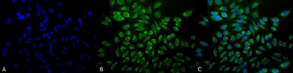
- Immunocytochemistry/Immunofluorescence analysis using human heat shocked cervical cancer cells. Fixation involved 2% Formaldehyde for 20 min at RT. Samples were incubated with GRP78 monoclonal antibody (Product # MA5-45279) at 1:100 for 12 hours at 4°C, followed by FITC Goat Anti-Mouse (green) at 1:200 for 2 hours at RT. Counterstain used was DAPI (blue) nuclear stain at 1:40,000 for 2 hours at RT. Localization: Endoplasmic reticulum lumen. Melanosome. Magnification: 20x. (A) DAPI (blue) nuclear stain. (B) Anti-GRP78 antibody. (C) Composite. Heat Shocked at 42°C for 1h.