 Explore
 Validate
 Learn
Learn Western blot
Antibody data
- Antibody Data
- Antigen structure
- References [0]
- Comments [0]
- Validations
- Western blot [2]
- Immunocytochemistry [1]
- Other assay [1]
Submit
Validation data
Reference
Comment
Report error
- Product number
- MA1-24631 - Provider product page

- Provider
- Invitrogen Antibodies
- Product name
- hnRNP C1/C2 Monoclonal Antibody (4F4)
- Antibody type
- Monoclonal
- Antigen
- Other
- Description
- Recommended positive controls: HeLa nuclear extract.
- Antibody clone number
- 4F4
- Concentration
- 2.4 mg/mL
No comments: Submit comment
Supportive validation
- Submitted by
- Invitrogen Antibodies (provider)
- Main image

- Experimental details
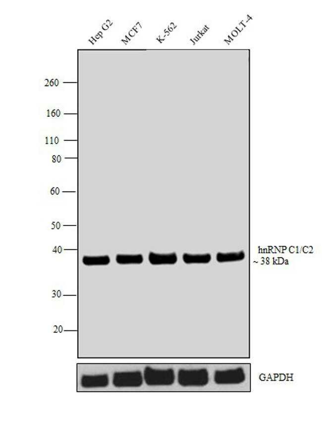
- Western blot analysis was performed on whole cell extracts (30 µg lysate) of HepG2 (Lane 1), MCF7 (Lane 2), K-562 (Lane 3), Jurkat (Lane 4) and MOLT-4 (Lane 5). The blot was probed with hnRNP C1/C2 Monoclonal Antibody (4F4) (Product # MA1-24631, 1:1000 dilution) and detected by chemiluminescence using Goat anti-Mouse IgG (H+L) Superclonal™ Secondary Antibody, HRP conjugate (Product # A28177, 0.25 µg/ml, 1:4000 dilution). A band at ~38 kDa corresponding to hnRNP C1/C2 was observed across all the cell lines tested.
- Submitted by
- Invitrogen Antibodies (provider)
- Main image
- Experimental details
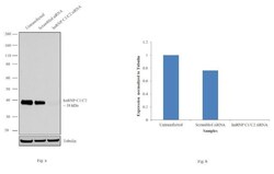
- Knockdown of hnRNP C was achieved by transfecting HeLa cells with hnRNP C specific siRNAs (Silencer® select Product # s6719, s6720). Western blot analysis (Fig. a) was performed using whole cell extracts from the hnRNP C knockdown cells (lane 3), non-specific scrambled siRNA transfected cells (lane 2) and untransfected cells (lane 1). The blots were probed with hnRNP C1/C2 Monoclonal Antibody (4F4) (Product # MA1-24631, 1:1000 dilution) and Goat anti-Mouse IgG (H+L) Superclonal™ Secondary Antibody, HRP conjugate (Product # A28177, 0.25 µg/ml, 1:4000 dilution). Densitometric analysis of this western blot is shown in histogram (Fig. b). Decrease in signal upon siRNA mediated knock down confirms that antibody is specific to hnRNP C.

Supportive validation
- Submitted by
- Invitrogen Antibodies (provider)
- Main image
- Experimental details
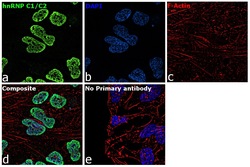
- Immunofluorescence analysis of hnRNP C1/C2 was performed using 70% confluent log phase HeLa cells. The cells were fixed with 4% paraformaldehyde for 10 minutes, permeabilized with 0.1% Triton™ X-100 for 15 minutes, and blocked with 1% BSA for 1 hour at room temperature. The cells were labeled with hnRNP C1/C2 Mouse Monoclonal Antibody (Product # MA1-24631) at 5 µg/mL in 0.1% BSA, incubated at 4 degree Celsius overnight and then labeled with Goat anti-Mouse IgG (H+L) Superclonal™ Secondary Antibody, Alexa Fluor® 488 conjugate (Product # A28175) at a dilution of 1:2000 for 45 minutes at room temperature (Panel a: green). Nuclei (Panel b: blue) were stained with ProLong™ Diamond Antifade Mountant with DAPI (Product # P36962). F-actin (Panel c: red) was stained with Rhodamine Phalloidin (Product # R415, 1:300). Panel d represents the merged image showing nucleus localization. Panel e represents control cells with no primary antibody to assess background. The images were captured at 60X magnification.

Supportive validation
- Submitted by
- Invitrogen Antibodies (provider)
- Main image

- Experimental details
- Detection of binding of endogenous hnRNP C1/C2 protein to specific RNA using Anti-hnRNP C1/C2 Antibody: RNA Immunoprecipitation (RIP) was performed using Anti-hnRNP C1/C2 Mouse Monoclonal Antibody (Product # MA1-24631, 5 µg) on clarified whole cell lysate from 4 million HCT 116 cells. Normal Rabbit IgG was used as a negative IP control. Immunoprecipitated RNA was purified by RiboPure™ RNA Purification Kit (Product # AM1924) and analyzed by RT-PCR using the Power SYBR® Green RNA-to-CT™ 1-Step Kit (Product # 4389986) with primer pairs over EGR1, ZFP36, ATF3, CYR61 (positive) and 18S rRNA (negative). Data is presented as fold enrichment of the antibody signal versus the negative control IgG using the comparative CT method.
