 Explore
 Validate
 Learn
Learn Western blot
Antibody data
- Antibody Data
- Antigen structure
- References [0]
- Comments [0]
- Validations
- Western blot [2]
- Immunocytochemistry [2]
- Immunoprecipitation [2]
- Immunohistochemistry [4]
- Other assay [1]
Submit
Validation data
Reference
Comment
Report error
- Product number
- PA5-117991 - Provider product page

- Provider
- Invitrogen Antibodies
- Product name
- hnRNP C Polyclonal Antibody
- Antibody type
- Polyclonal
- Antigen
- Other
- Reactivity
- Human
- Host
- Rabbit
- Isotype
- IgG
- Vial size
- 100 μL
- Storage
- -20°C, Avoid Freeze/Thaw Cycles
No comments: Submit comment
Supportive validation
- Submitted by
- Invitrogen Antibodies (provider)
- Main image

- Experimental details
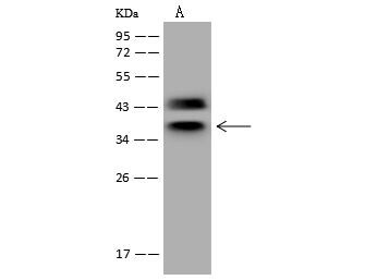
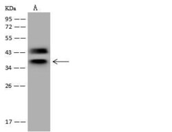
- Western Blot using hnRNP C Polyclonal Antibody (Product # PA5-117991) at 1:500 dilution. Lane A: HeLa Whole Cell Lysate. Lysates/proteins at 30 μg per lane. Secondary antibody: Goat Anti-Rabbit IgG (H+L)/HRP at 1:10,000 dilution. Developed using the ECL technique. Performed under reducing conditions. Predicted band size: 34 kDa. Observed band size: 36 kDa.
- Submitted by
- Invitrogen Antibodies (provider)
- Main image
- Experimental details
- Western Blot of hnRNP C in Lane A: HeLa Whole Cell Lysate. Samples (30 µg per lane) were incubated with polyclonal antibody (Product # PA5-117991) with a dilution of 1:500 , followed by Goat Anti-Rabbit IgG (H+L)/HRP using a dilution of 1:10,000. Assay was performed under reducing conditions. Predicted band size: 34 kDa, Observed band size: 36 kDa.
Supportive validation
- Submitted by
- Invitrogen Antibodies (provider)
- Main image
- Experimental details
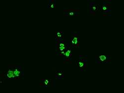
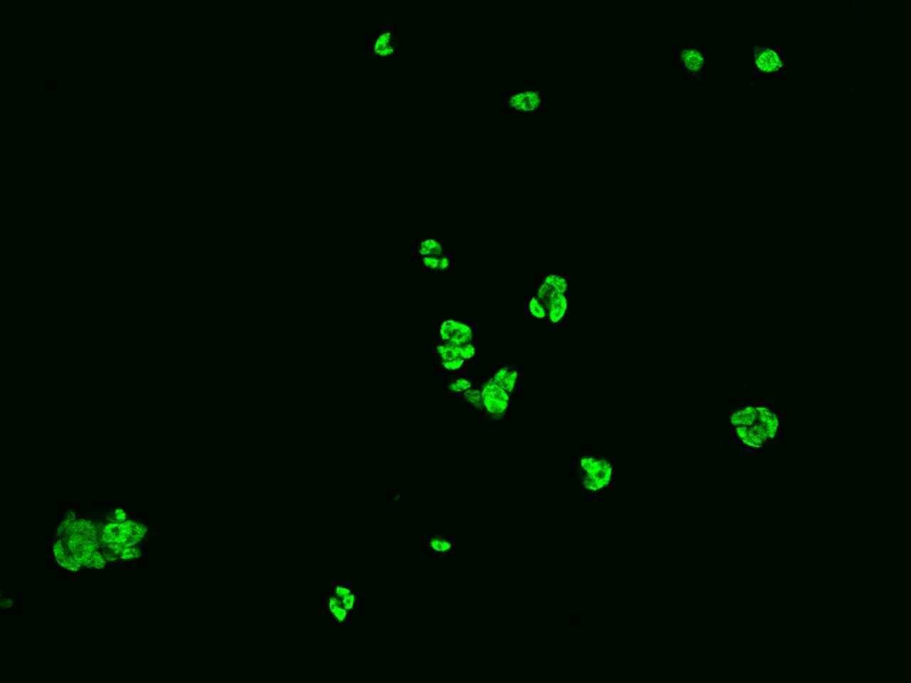
- Immunofluorescence staining of hnRNP C in HEK293 cells. Cells were fixed with 4% PFA, permeabilzed with 0.1% Triton X-100 in PBS, blocked with 10% serum, and incubated with hnRNP C Polyclonal Antibody (Product # PA5-117991, 1:200) at 4°C overnight. Then cells were stained with the Alexa Fluor®488-conjugated Goat Anti-rabbit IgG secondary antibody (green). Positive staining was localized to nucleus.
- Submitted by
- Invitrogen Antibodies (provider)
- Main image

- Experimental details
- Immunofluorescence staining of hnRNP C in HEK293 cells. Cells were fixed with 4% PFA, permeabilzed with 0.1% Triton X-100 in PBS, blocked with 10% serum, and incubated with hnRNP C Polyclonal Antibody (Product # PA5-117991, 1:200) at 4°C overnight. Then cells were stained with the Alexa Fluor®488-conjugated Goat Anti-rabbit IgG secondary antibody (green). Positive staining was localized to nucleus.

Supportive validation
- Submitted by
- Invitrogen Antibodies (provider)
- Main image
- Experimental details
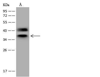
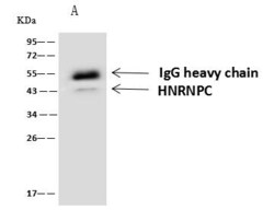
- hnRNP C Immunoprecipitation using: Lane A: 0.5 mg HeLa Whole Cell Lysate 4 µL with hnRNP C Polyclonal Antibody (Product # PA5-117991) and 60 μg of Immunomagnetic beads Protein A/G. Primary antibody: hnRNP C Polyclonal Antibody, at 1:100 dilution. Secondary antibody: Goat Anti-Rabbit IgG (H+L) /HRP at 1:10,000 dilution. Developed using the ECL technique. Performed under reducing conditions. Predicted band size: 34 kDa. Observed band size: 43 kDa.

- Submitted by
- Invitrogen Antibodies (provider)
- Main image
- Experimental details
- Immunoprecipitation of hnRNP C in Lane A: 0.5 mg HeLa Whole Cell Lysate. Samples were treated with 60 μg of immunomagnetic Protein A/G beads, incubated with polyclonal antibody (Product # PA5-117991) with a dilution of 1:100 , followed by Goat Anti-Rabbit IgG (H+L)/HRP using a dilution of 1:10,000. Assay was performed under reducing conditions. Predicted band size: 34 kDa , Observed band size : 43 kDa .

Supportive validation
- Submitted by
- Invitrogen Antibodies (provider)
- Main image
- Experimental details
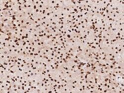
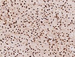
- Immunohistochemistry (Paraffin) of hnRNP C in human liver. Samples were incubated with polyclonal antibody (Product # PA5-117991) with a dilution of 1:100 .

- Submitted by
- Invitrogen Antibodies (provider)
- Main image
- Experimental details
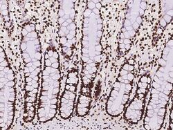
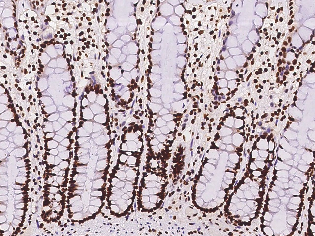
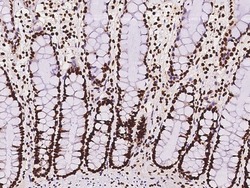
- Immunohistochemical staining of human hnRNP C in human rectum with hnRNP C Polyclonal Antibody (Product # PA5-117991, 1:100 dilution, formalin-fixed paraffin embedded sections).
- Submitted by
- Invitrogen Antibodies (provider)
- Main image
- Experimental details
- Immunohistochemistry (Paraffin) of hnRNP C in human rectum. Samples were incubated with polyclonal antibody (Product # PA5-117991) with a dilution of 1:100 .

- Submitted by
- Invitrogen Antibodies (provider)
- Main image
- Experimental details
- Immunohistochemistry (Paraffin) of hnRNP C in human liver. Samples were incubated with polyclonal antibody (Product # PA5-117991) with a dilution of 1:100 .

Supportive validation
- Submitted by
- Invitrogen Antibodies (provider)
- Main image

- Experimental details
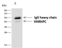
- hnRNP C Immunoprecipitation using: Lane A: 0.5 mg HeLa Whole Cell Lysate 4 µL with hnRNP C Polyclonal Antibody (Product # PA5-117991) and 60 μg of Immunomagnetic beads Protein A/G. Primary antibody: hnRNP C Polyclonal Antibody, at 1:100 dilution. Secondary antibody: Goat Anti-Rabbit IgG (H+L) /HRP at 1:10,000 dilution. Developed using the ECL technique. Performed under reducing conditions. Predicted band size: 34 kDa. Observed band size: 43 kDa.
