 Explore
 Validate
 Learn
Learn Western blot
Antibody data
- Antibody Data
- Antigen structure
- References [1]
- Comments [0]
- Validations
- Western blot [6]
- Immunocytochemistry [2]
- Immunohistochemistry [2]
Submit
Validation data
Reference
Comment
Report error
- Product number
- PA5-22280 - Provider product page

- Provider
- Invitrogen Antibodies
- Product name
- hnRNP C Polyclonal Antibody
- Antibody type
- Polyclonal
- Antigen
- Recombinant protein fragment
- Description
- Recommended positive controls: Raji, Neuro2A, NIH-3T3, BCL-1, Raw264.7, C2C12, PC-12, Rat2.
- Concentration
- 0.79 mg/mL
Submitted references Circ_0006790 carried by bone marrow mesenchymal stem cell-derived exosomes regulates S100A11 DNA methylation through binding to CBX7 in pancreatic ductal adenocarcinoma.
Gao G, Wang L, Li C
American journal of cancer research 2022;12(5):1934-1959
American journal of cancer research 2022;12(5):1934-1959
No comments: Submit comment
Supportive validation
- Submitted by
- Invitrogen Antibodies (provider)
- Main image
- Experimental details
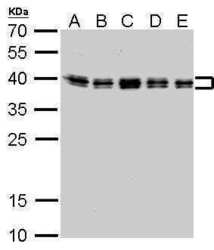
- hnRNP C Polyclonal Antibody detects HNRNPC protein by western blot analysis. A. 30 µg Neuro2A whole cell lysate/extract. B. 30 µg NIH-3T3 whole cell lysate/extract. C. 30 µg BCL-1 whole cell lysate/extract. D. 30 µg Raw264.7 whole cell lysate/extract. E. 30 µg C2C12 whole cell lysate/extract.12% SDS-PAGE. HnRNP C Polyclonal Antibody (Product # PA5-22280) dilution: 1:1,000. The HRP-conjugated anti-rabbit IgG antibody was used to detect the primary antibody.

- Submitted by
- Invitrogen Antibodies (provider)
- Main image

- Experimental details
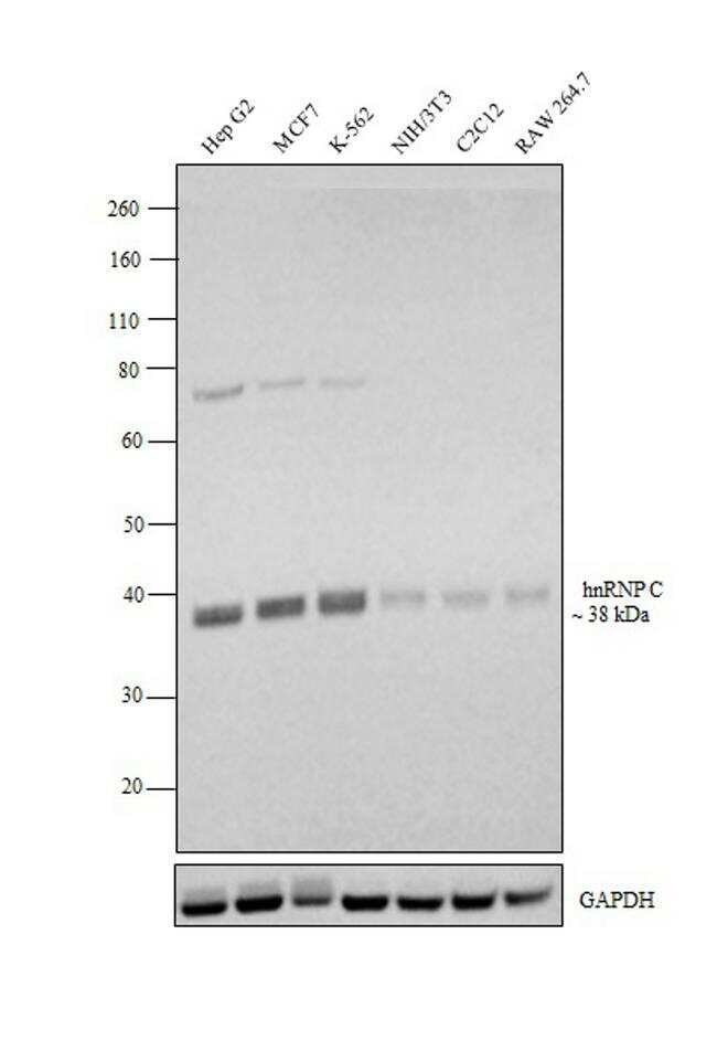
- Western blot analysis was performed on whole cell extracts (30 µg lysate) of HepG2 (Lane 1), MCF7 (Lane 2), K-562 (Lane 3), NIH/3T3 (Lane 4), C2C12 (Lane 5) and Raw 264.7 (Lane 6). The blot was probed with hnRNP C Polyclonal Antibody (Product # PA5-22280, 1:1000 dilution) and detected by chemiluminescence using Goat anti-Rabbit IgG (H+L) Superclonal™ Secondary Antibody, HRP conjugate (Product # A27036, 0.25 µg/ml, 1:4000 dilution). A band at ~38 kDa corresponding to hnRNP C was observed across all the cell lines tested.
- Submitted by
- Invitrogen Antibodies (provider)
- Main image
- Experimental details
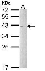
- Western Blot using hnRNP C Polyclonal Antibody (Product # PA5-22280). Sample (30 µg of whole cell lysate). Lane A: Raji. 12% SDS PAGE. hnRNP C Polyclonal Antibody (Product # PA5-22280) diluted at 1:1,000. The HRP-conjugated anti-rabbit IgG antibody was used to detect the primary antibody.

- Submitted by
- Invitrogen Antibodies (provider)
- Main image

- Experimental details
- hnRNP C Polyclonal Antibody detects HNRNPC protein by western blot analysis. A. 30 µg Neuro2A whole cell lysate/extract. B. 30 µg NIH-3T3 whole cell lysate/extract. C. 30 µg BCL-1 whole cell lysate/extract. D. 30 µg Raw264.7 whole cell lysate/extract. E. 30 µg C2C12 whole cell lysate/extract.12% SDS-PAGE. HnRNP C Polyclonal Antibody (Product # PA5-22280) dilution: 1:1,000. The HRP-conjugated anti-rabbit IgG antibody was used to detect the primary antibody.

- Submitted by
- Invitrogen Antibodies (provider)
- Main image

- Experimental details
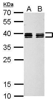
- hnRNP C Polyclonal Antibody detects HNRNPC protein by western blot analysis. A. 30 µg PC-12 whole cell lysate/extract. B. 30 µg Rat2 whole cell lysate/extract.12% SDS-PAGE. HnRNP C Polyclonal Antibody (Product # PA5-22280) dilution: 1:1,000. The HRP-conjugated anti-rabbit IgG antibody was used to detect the primary antibody.
- Submitted by
- Invitrogen Antibodies (provider)
- Main image

- Experimental details
- Knockdown of hnRNP C was achieved by transfecting HeLa cells with hnRNP C specific siRNAs (Silencer® select Product # s6719, s6720). Western blot analysis (Fig. a) was performed using whole cell extracts from the hnRNP C knockdown cells (lane 3), non-specific scrambled siRNA transfected cells (lane 2) and untransfected cells (lane 1). The blots were probed with hnRNP C Polyclonal Antibody (Product # PA5-22280, 1:1000 dilution) and Goat anti-Rabbit IgG (H+L) Superclonal™ Secondary Antibody, HRP conjugate (Product # A27036, 0.25 µg/ml, 1:4000 dilution). Densitometric analysis of this western blot is shown in histogram (Fig. b). Decrease in signal upon siRNA mediated knock down confirms that antibody is specific to hnRNP C.

Supportive validation
- Submitted by
- Invitrogen Antibodies (provider)
- Main image
- Experimental details
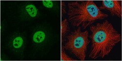
- Immunocytochemistry-Immunofluorescence analysis of hnRNP C was performed in HeLa cells fixed in 4% paraformaldehyde at RT for 15 min. Green: hnRNP C Polyclonal Antibody (Product # PA5 22280) diluted at 1:500. Red: alpha Tubulin, a cytoskeleton marker. Blue: Hoechst 33342 staining.

- Submitted by
- Invitrogen Antibodies (provider)
- Main image

- Experimental details
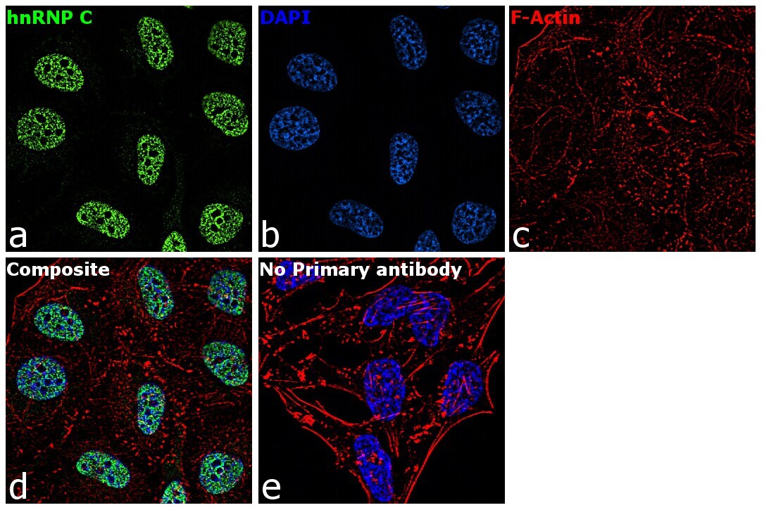
- Immunofluorescence analysis of hnRNP C was performed using 70% confluent log phase HeLa cells. The cells were fixed with 4% paraformaldehyde for 10 minutes, permeabilized with 0.1% Triton™ X-100 for 15 minutes, and blocked with 1% BSA for 1 hour at room temperature. The cells were labeled with hnRNP C Rabbit Polyclonal Antibody(Product # PA5-22280) at 5 µg/mL in 0.1% BSA, incubated at 4 degree Celsius overnight and then labeled with Goat anti-Rabbit IgG (H+L) Superclonal™ Secondary Antibody, Alexa Fluor® 488 conjugate (Product # A27034) at a dilution of 1:2000 for 45 minutes at room temperature (Panel a: green). Nuclei (Panel b: blue) were stained with ProLong™ Diamond Antifade Mountant with DAPI (Product # P36962). F-actin (Panel c: red) was stained with Rhodamine Phalloidin (Product # R415, 1:300). Panel d represents the merged image showing nucleus localization. Panel e represents control cells with no primary antibody to assess background. The images were captured at 60X magnification.
Supportive validation
- Submitted by
- Invitrogen Antibodies (provider)
- Main image
- Experimental details
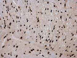
- Immunohistochemistry (Paraffin) analysis of hnRNP C was performed in paraffin-embedded mouse brain tissue using hnRNP C Polyclonal Antibody (Product # PA5-22280) at a dilution of 1:500.

- Submitted by
- Invitrogen Antibodies (provider)
- Main image
- Experimental details
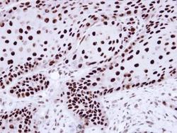
- Immunohistochemical analysis of paraffin-embedded Cal27 Xenograft, using hnRNP C1/C2 (Product # PA5-22280) antibody at 1:100 dilution. Antigen Retrieval: Citrate buffer, pH 6.0, 15 min.
