Explore
 Validate
 Learn
Learn Western blot
Antibody data
- Antibody Data
- Antigen structure
- References [1]
- Comments [0]
- Validations
- Western blot [1]
- Immunohistochemistry [1]
- Other assay [1]
Submit
Validation data
Reference
Comment
Report error
- Product number
- PA5-79323 - Provider product page

- Provider
- Invitrogen Antibodies
- Product name
- GRB7 Polyclonal Antibody
- Antibody type
- Polyclonal
- Antigen
- Synthetic peptide
- Description
- Reconstitute with 0.2 mL of distilled water to yield a concentration of 500 µg/mL. Positive Control - WB: Rat Testis Tissue, SMMC whole cell, HELA whole cell, A549 whole cell, SW620 whole cell. IHC: Human Oesophagus Squama Cancer Tissue.
- Reactivity
- Human, Mouse, Rat
- Host
- Rabbit
- Isotype
- IgG
- Vial size
- 100 μg
- Concentration
- 500 μg/mL
- Storage
- -20°C
Submitted references Focal adhesion ribonucleoprotein complex proteins are major humoral cancer antigens and targets in autoimmune diseases.
Atsumi S, Katoh H, Komura D, Hashimoto I, Furuya G, Koda H, Konishi H, Suzuki R, Yamamoto A, Yuba S, Abe H, Rino Y, Oshima T, Ushiku T, Fukayama M, Seto Y, Ishikawa S
Communications biology 2020 Oct 16;3(1):588
Communications biology 2020 Oct 16;3(1):588
No comments: Submit comment
Supportive validation
- Submitted by
- Invitrogen Antibodies (provider)
- Main image
- Experimental details
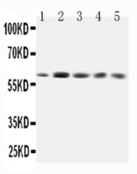
- Western blot analysis of GRB7 in Lane 1: rat testis tissue lysate, Lane 2: SMMC whole cell lysate, Lane 3: HeLa whole cell lysate, Lane 4: A549 whole cell lysate, Lane 5: SW620 whole cell lysate using 40 µg per well. Sample was incubated with GRB7 (Product # PA5-79323) at a dilution of 0.5 µg/mL.

Supportive validation
- Submitted by
- Invitrogen Antibodies (provider)
- Main image
- Experimental details
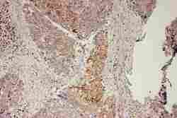
- Immunohistochemistry analysis of GRB7 on paraffin-embedded human Esophagus Squama cancer tissue. Sample was incubated with GRB7 polyclonal antibody (Product# PA5-79323).
Supportive validation
- Submitted by
- Invitrogen Antibodies (provider)
- Main image
- Experimental details
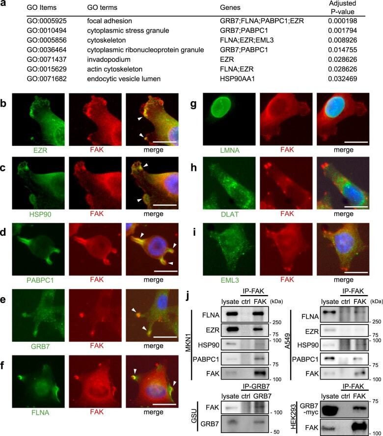
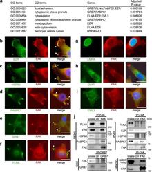
- Fig. 3 Focal adhesion-related protein complexes are major and common humoral antigens in tumor microenvironments. a GO enrichment analysis of the eight identified protein antigens. b - i Fluorescent immunocytochemistry of the identified protein antigens (green) along with focal adhesion kinase (FAK) (red) onto a human gastric cancer cell line MKN1. The blue color represents Hoechst nuclear staining. The white bars indicate 20 mum. The white arrowheads indicate representative hot spots of colocalization between protein antigens and focal adhesions (only in b - f ). Similar colocalizations were reproducibly observed in all analyzed cells with formations of visible focal adhesions (only in b - f ). The same immunocytochemical staining was also performed onto another human gastric cancer cell line GSU, obtaining similar results (Supplementary Fig. 4 ). j Co-immunoprecipitation experiments of the FAK complex. The upper and lower panels at the left side show co-IP using anti-FAK antibody for MKN1 and anti-GRB7 antibody for GSU, respectively, followed by immunoblots using the indicated antibodies. The right-side panels show co-IP using anti-FAK antibody for A549, a lung adenocarcinoma cell line, and HEK293 transfected with GRB7-myc/His construct, respectively, followed by immunoblots using the indicated antibodies. Co-IP experiments were conducted at least twice.